Medical Applications
Medical applications of physics investigated in the Department include the development of novel imaging techniques, work on detecting and treating cancer, the use of nanomaterials and techniques, and the development of AI-based tools for medicine.
Medical imaging
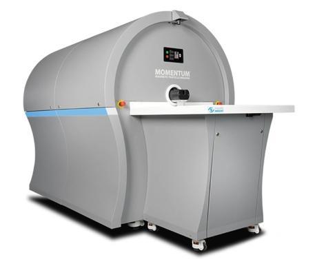
Medical imaging methods based on magnetic nanoparticles offer many advantages over MRI, including higher speed and lower cost. Work in Physics, in collaboration with the Chemistry Department and the Medical Faculty, has led to the UK’s first pre-clinical Magnetic Particle Imaging system being established in Liverpool.
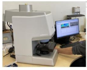
The Department has pioneered the use of Fourier Transform Infra-Rad (FTIR) imaging, coupled with AI image-analysis, to identify oral cancer. The method has been patented and development of a clinical system based on the technique is in progress.
Proton and ion therapy
The Liverpool region has played host to the UK’s first proton therapy machine, the cyclotron at Clatterbridge Cancer Centre dedicated to the treatment of okular tumours. The Department has a long-standing interest in hadron therapy for cancer treatment; current projects include the development of non-invasive methods of monitoring proton and ion therapy beams (at the Cockcroft Institute), and of advanced, tissue-equivalent instrumented phantoms (with FBK, Trento), which allow precise monitoring of the dose delivered in proton and ion therapy.

Artificial intelligence in medicine
In addition to the uses of AI mentioned above, further applications include the development of models for the diagnosis of age-related macular degeneration and diabetic retinopathy.
Safety critical applications of AI, such as those in medicine, raise concerns because of the “black-box” nature of many AI systems. To address this problem, Liverpool is leading an EU-funded international consortium investigating the use of explainable AI methods.
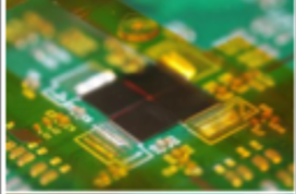
Development of use of gamma-ray detectors
A further strand of work that has long been a feature of research at Liverpool is the development of highly sensitive gamma-ray detectors for medical use, for example CZT-based imaging systems for low dose breast imaging and for dosimetry during radionuclide therapy. Imaging of the prompt gamma-rays produced during proton beam radiotherapy, which has the potential to provide real-time information on the dose a patient receives, has also been investigated.