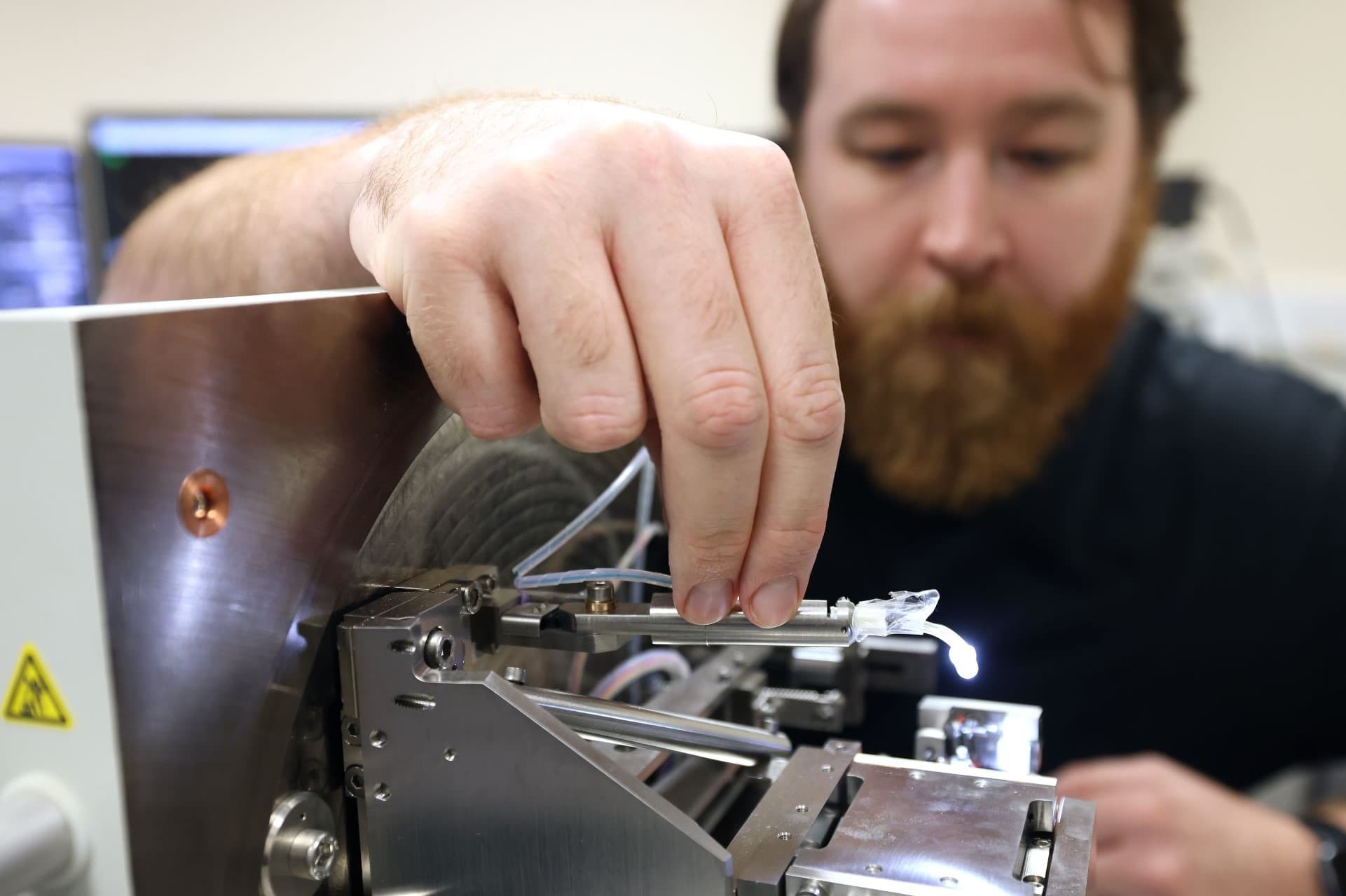
The Electron Microscopy Facility is fully equipped for transmission electron microscopy (TEM), scanning electron microscopy (SEM), and serial block faces SEM (SBF-SEM). We are specialists in correlative light-electron microscopy (CLEM) and volumeCLEM. We also work with our colleagues in the Faculty of Science and Engineering to provide seamless multi-modal workflows.
The facility is supported by expert academic and research technical professionals, who can provide training and support in all aspects of sample preparation, imaging, image analysis and interpretation. The facility has an open access approach to internal and external researchers across biological, chemical, and material sciences.
We operate a full service, independent or collaborative model of access and we are always happy to discuss individual research requirements.
What you'll receive from our facility
- Our facility staff are actively engaged with the microscopy community, both nationally and internationally, to keep up to date with imaging trends and techniques
- We work across both Electron Microscopy and Light Microscopy platforms to provide smooth CLEM workflows
- Staff are available to share their expertise in imaging a diverse range of biological samples from proteins, viruses, phages, cells, organoids through to tissues, with tried and tested protocols already optimised for many experimental conditions
- We are able to provide support from initial project discussions and planning through to training, experimental techniques and assistance with data analysis and interpretation
- Support is also available with grant applications, method development, data management planning, and costings
- Initial training and ongoing support are available throughout the lifetime of a project.
The equipment we offer includes:
- 120Kv Transmission Electron Microscope
- FEG Scanning Electron Microscope
- SBF-SEM (3D-vEM)
- Benchtop Scanning Electron Microscope
- Sample preparation equipment.
Who can use our facilities
- University of Liverpool academic, research and technical staff
- Researchers from other universities/research institutions
- Industrial research partners
- Clinicians/surgeons.
What Biomedical Electron Microscopy can be used for
Although the facility specialises in the analysis of room temperature biological samples, we can produce high-resolution imaging of many other types of samples and materials. All our systems can be used for a variety of applications, meaning we can offer processing and imaging options to suit your research objectives.
Our other capabilities include:
- Negative staining of biological samples
- ‘Rip Off’ labelling of cell membrane sheets
- Cryo-EM screening
- RT 3D tomography
- Hydrated samples in ESEM mode
- Pre and post embedding Immunogold labelling
- Serial section TEM
- Cryo-sectioning and labelling – Tokuyasu method.
We are fully equipped for safe reagent preparation including making gold probes and plastic/carbon coated TEM grids.


