The Centre for Pre-Clinical Imaging offers researchers state-of-the-art technologies for multi-modality, non-invasive approaches to imaging on small, live animals (in vivo), or tissues and organs extracted from animals (ex vivo). Our equipment can be used for pre-clinical models and non-biological samples.
The Facility’s next-generation equipment and resources are supported by experienced and knowledgeable technical specialists and renowned academics to help you make the most of your data.
What you'll receive from our facility
- Access to pre-clinical MRI, small animal µCT, PET/SPECT/CT imaging, small animal ultrasound, bioluminescence/fluorescence imaging (IVIS) and small animal optoacoustic imaging
- Data processing workstations running a range of proprietary and open-source software for image processing and analysis
- Expert training and ongoing support for imaging systems in CPI. Expert support for data processing and analysis
- Support for grant applications, manuscript preparation and costings
- Bespoke method development for novel imaging applications.
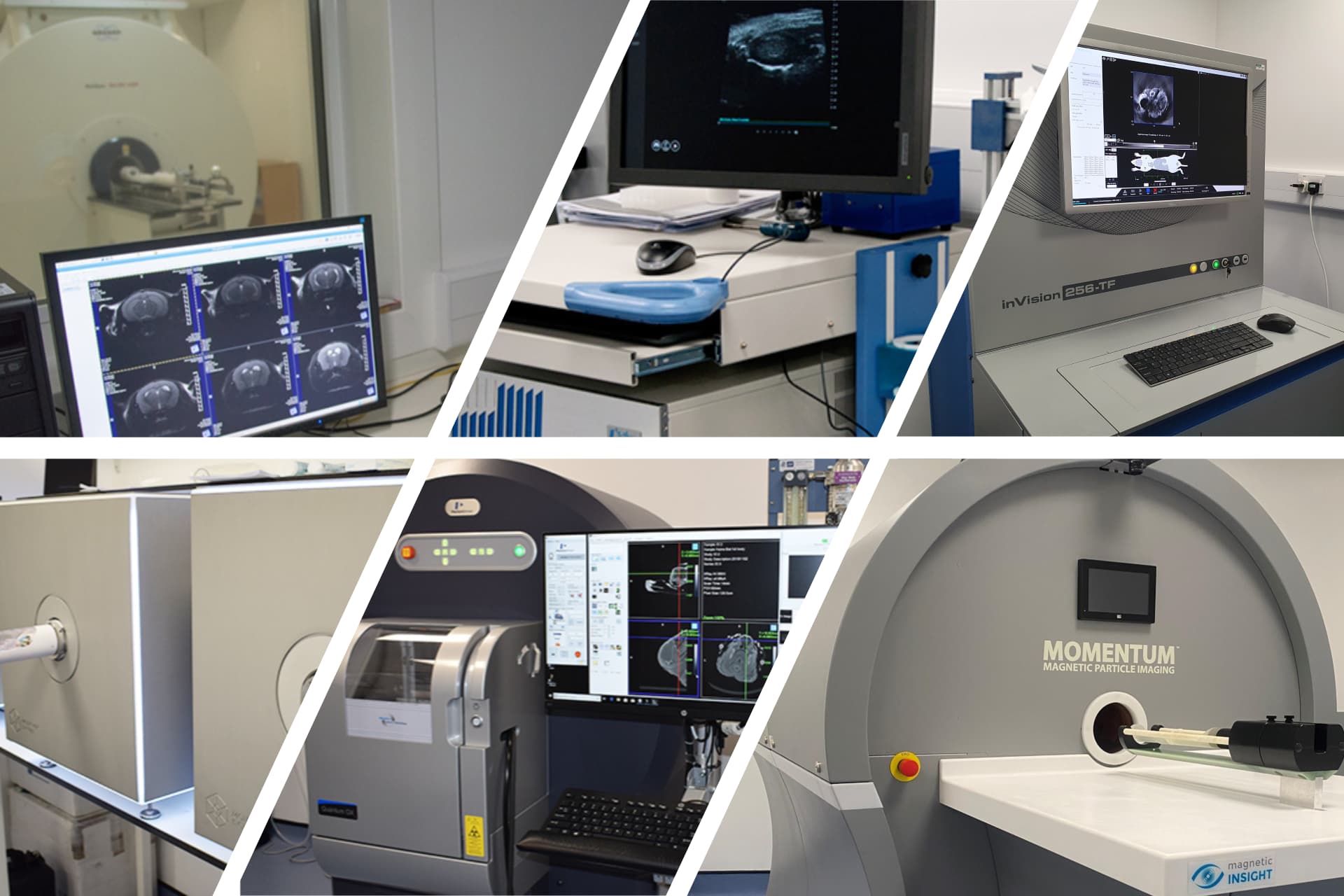
The equipment we offer includes:
- Magnetic Resonance Imaging and Spectroscopy (Bruker, 9.4 T scanner)
- Bioluminescence & Fluorescence imaging (PerkinElmer, IVIS)
- Ultrasound imaging (S-Sharp, Prospect T1)
- Optoacoustic imaging (iThera Medical, MSOT-Invision 256-TF)
- µCT imaging (PerkinElmer, Quantum GX2)
- PET+SPECT+CT imaging systems (Molecubes, β+ɣ+X cubes)
- Magnetic Particle Imaging (Magnetic Insight, Momentum)
- Image processing software (both licensed and open source).
We work towards maintaining the highest standards of animal welfare. Our ethical review process involves individuals from a variety of backgrounds, including vets, animal welfare officers, scientists and lay people.
See the University of Liverpool's animal research policy.
Who can use our facilities?
- University of Liverpool academic staff
- Researchers from other universities
- Industrial research partners.
All researchers - University of Liverpool based, external and industrial research partners can use our services to test out if a drug, procedure, or treatment is likely to be useful.
University of Liverpool academic staff can apply for research funding vouchers.
What Pre-Clinical Imaging can be used for
We provide users with the ability to carry out non-invasive, longitudinal, high-resolution imaging of small animals. This allows measurement of physiological or metabolic response to disease or drug treatment. Amongst other things, we can non-invasively image anatomy as well as directly measure in vivo metabolism, carry out cell tracking, measure cardiac function, all without injury to the animal. We can combine multiple imaging technologies enabling multimodal imaging. Non-invasive longitudinal imaging helps reduce animal numbers required for a study, and aids with 3Rs compliance.
Whilst our scanners are optimized for small animal work, they are general-purpose imaging scanners and are equally well suited to imaging ex vivo and non-biological samples. We can carry out high-resolution imaging of individual organs or post-mortem samples as well as materials (e.g. µCT imaging of microcircuitry).
Our team can develop new bespoke imaging protocols, methods and hardware to help you get the data that you need.