Evolutionary Morphology and Biomechanics Group
Ageing is associated with several conditions which affect the mechanics of the human musculoskeletal system, including diabetes and osteoarthritis, and decline in cognitive and locomotor capabilities greatly increase risk of falling. Our companion animals, too, are subject to conditions which affect their ability to move normally, some the result of breed standards which focus on desired physical attributes rather than wellbeing and health. We are addressing each of these, and even the effects of environmental factors such as substrate types and footwear design.
Evolutionary Morphology and Biomechanics Group
About our Research
Our research focuses on musculoskeletal functioning at the whole-body level. By doing so, we build on the expertise of our colleagues at the Institute of Life Course and Medical Sciences who focus on underlying organisational levels, from molecules to tissues and organs.
We work on humans but also on other animals - either as models or for their own merit. We strongly believe that, in many cases, a comparative and evolutionary approach is necessary to help us understand how healthy bodies work. Such fundamental insight is required to fully understand (dis)functioning, in particular as a result of ageing and chronic disease. We study a variety of topics from fundamental to more applied. We have listed a few examples, below.
Our group uses experimental and modelling approaches as appropriate. Our gait lab is fully equipped with motion capture systems, force plates and additional techniques including electromyography, inertial sensors and isokinetic muscle testing. We also house the CIMA/MRC biplanar X-Ray facilities. For modelling, we use 3D shape reconstruction based on medical imaging and a variety of modelling techniques (including link-segment, finite element and evolutionary robotics).
Please see our lab’s blog at embliverpool.wordpress.com for more information.
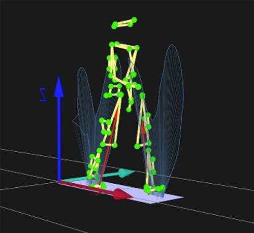
Figure 1: Quantifying human gait by measuring kinematics (motions, see the green markers) and kinetics (forces, see the red ground reaction force vectors).
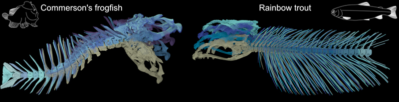
Figure 2: Revealing how fish create neck-like motions and 3D spine mobility, to develop better models of 3D musculoskeletal dynamics in during healthy performance and ageing.
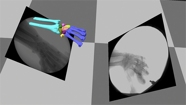
Figure 3: In collaboration with the School of Engineering and surgeons from Liverpool Hospitals, we are using a cadaveric robotic simulator in conjunction with biplanar x-ray videography to investigate normal wrist motion and how best to restore healthy motion through surgical interventions necessitated by injury or age-related disease.
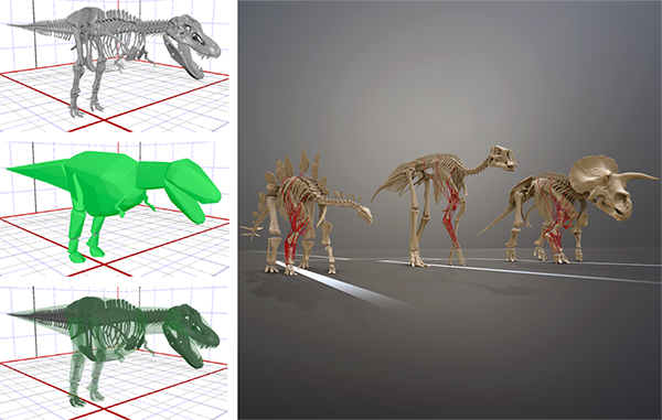
Figure 4: We combine 3D digitisation and computational approaches with quantitative soft tissue data from living animals to reconstruct musculoskeletal anatomy and limb biomechanics in extinct animals like dinosaurs.
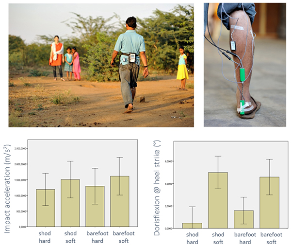
Figure 5: We work in field settings as well as laboratory settings. This example shows field work quantifying gait on natural substrates using indigenous footwear in South India (top). Data show differences in heel impact acceleration and foot posture at heel strike between substrate and footwear conditions.
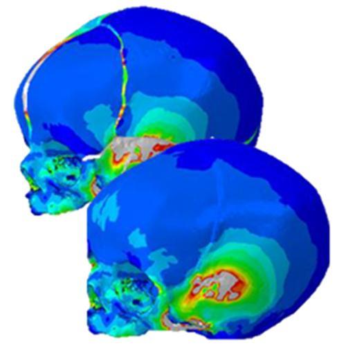
Figure 6: Finite element analysis of infant brain expansion and muscle activation shows the importance of cranial sutures for healthy skull growth.
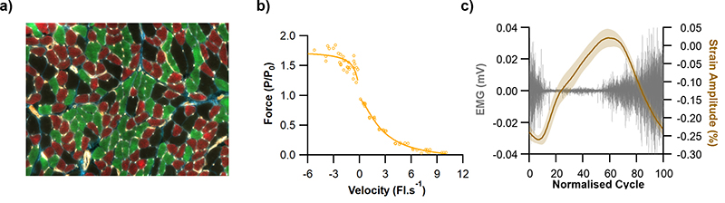
Figure 7: From fibre to function: understanding how muscle structure underpins muscle function. Our approach tries to understand how muscle architecture (e.g. fibre length, muscle mass) or in this example muscle fibre type composition (a) links to gross muscle function (e.g. a muscles force velocity profile) (b). This structural (a) and isolated functional (b) information is then related to in vivo function of a muscle, for example combining recordings of joint kinematics with muscle activity (electromyography) and muscle fibre strain data (c). Understanding how muscle functions in normal non-pathological tissue is critical to be able to effectively evaluate muscle properties across disease populations and develop effective therapeutic treatment strategies. This approach is also vital in the development and validation of musculoskeletal models which rely on these physiological input parameters.


