Clinical presentation
The primary feature of BDD is lameness, most commonly involving (~80-90%) on the hind feet. Lameness may become non-weight bearing or animals may shift weight from foot to foot or walk on their toes.
This lameness is caused by a lesion which is usually located between the heel bulbs, immediately above the coronet, but may affect other sites, including the skin of interdigital cleft, heel or dew claw. Chronic lesions frequently develop wart-like papillary keratotic proliferations. Typically, lesions are painful, moist, malodorous and prone to bleeding. Lesions appear as a focally-inflamed ulcerative lesion of circumscribed, dark red or brown hyperkeratotic skin.
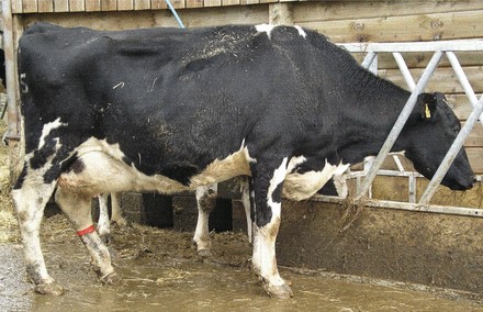
Lameness observed in a dairy cow with BDD
A now widely-adopted scoring system based on macroscopic lesion characteristics is shown in the table below. Lesions initially appear as small red/grey and circumscribed (M1) and typically increase in size to become large (>2cm) and intensely painful (M2). Following treatment, lesions will usually resolve (M3), although in untreated animals, lesions may become chronic (M4-M4.1). Importantly, there is a high level of disease recurrence in all animals, regardless of history.
| Grade | Macroscopic observations |
|---|---|
| M1 | A small focal active red/grey circumscribed lesion <2cm wide with 1mm wide red foci. |
| M2 | A painful large ulcerative, red/grey active lesion >cm wide. |
| M3 | A healing, painless brown scab; typically seen after treatment. |
| M4 | A chronic, stage presenting as dyskeratosis or irregular proliferative hyperkeratotic overgrowths. |
| M4.1 | A chronic stage with active painful M1 focus. |
| M5/M0 | Healthy skin with no evidence of previous lesion. |
The images below provide examples of active (M2) stage BDD lesions.
An M2 (active ulcerative) stage DD lesion, greater than 2 cm in diameter, with a red-grey mottled appearance.

An M2 (active ulcerative) stage DD lesion, greater than 2 cm in diameter, appearing very red and moist.
