Gamma scintigraphy imaging (bone scans)
Gamma scintigraphy, commonly known as ‘bone scan’, is a type of advanced imaging that may be indicated to further investigate specific types of problems in horses. We use the same type of radioactive substance (Technetium-99m) that is used in people to investigate specific types of bone abnormalities.
When are bone scans needed?
The most common reasons for performing these scans in horses admitted to the hospital are:
- Investigation of poor performance
- More complex lameness cases including horses that are lame in more than one leg (multi-limb lameness cases)
- Suspected fractures in areas that are difficult to assess in other areas of the body using more standard X-ray or ultrasound imaging e.g. pelvis
- Horses that will not tolerate routine lameness investigations involving nerve blocks.
What does a bone scan involve?
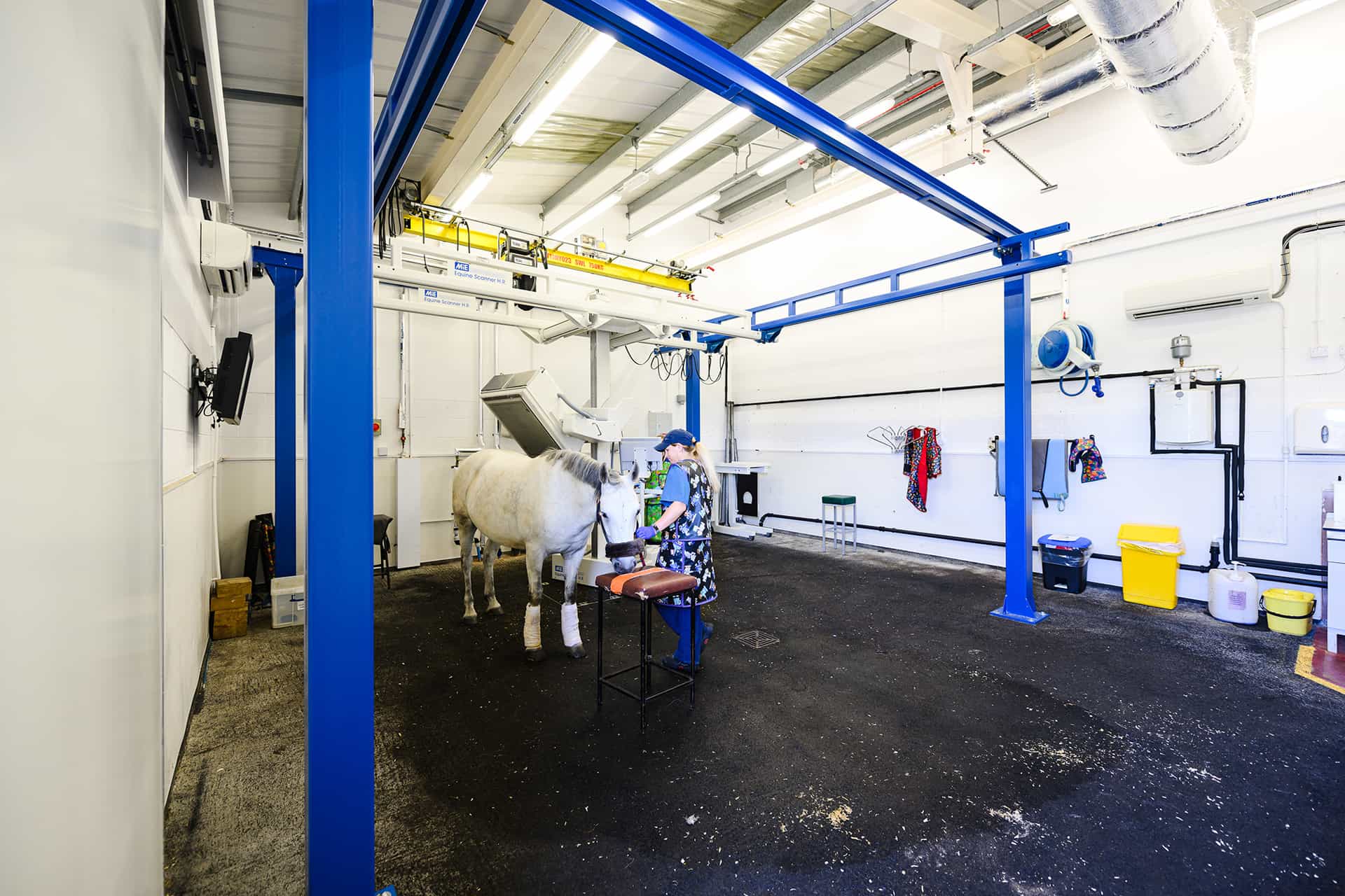
A bone scan involves the injection of a radioactive substance that attaches to areas of bone with areas of abnormal activity. A specialised camera is used to scan the bones we suspect are the underlying source of the problem to help locate these abnormal areas.
Horses undergoing bone scans are kept in our specifically designed radiation isolation stables while they are radioactive. The radioactive substance is administered by of our trained staff. Horses have an intravenous catheter placed into the jugular (neck) vein so that they do not need repeated injections. The radioactive substance takes only a few seconds to administer via the intravenous catheter and does not cause the horse any pain. This substance will then circulate around their body, reaching the areas of bone we need to image. Usually it takes 2-3 hours for this to happen.
The horse is then walked a short distance to our gamma scan unit for imaging using a specifically designed camera. The gamma scan itself is non-invasive, the camera is placed close to the horse at specific locations and angles. This is performed with the horse standing. However, to get the most diagnostic images, we need to remain still whilst each image is obtained, which can take around a minute each.
Therefore, horses are sedated to keep them relaxed and to make the scanning time as short as possible. The process of obtaining the scans itself takes around 2-3 hours depending on how many areas we need to image.
What happens after the bone scan?
Because horses remain radioactive for 48 hours after the injection, once the scans have been completed, they are returned to their radiation isolation stable. After the 48-hour period, they are then moved to one of our standard stable blocks.
The multiple images obtained are then assessed in-depth by members of our hospital team who are highly experienced in interpreting the scans. Sometimes, it is obvious where the area of abnormal bone and site of the problem is. However, some abnormal areas of uptake of the radioactive marker are not associated with the main source of pain or discomfort for the horse.
Once the scans have been assessed, we will discuss the findings and initial plan with the owner and their veterinary surgeon. This may involve further investigations focusing on the abnormal areas identified including nerve blocks, X-ray or ultrasound as appropriate or treatment involving medication or surgery.
This may be done at the hospital by our team of specialists or by the horse’s usual veterinary practice depending on the findings of the scan and the type of further investigations and treatments needed.