Diagnostic Radiography
UCAS code B821
- Study mode
- Full-time
- Duration
- 3 years
- Start date and application deadlines
-
- Start date
- September 2026
- Apply by:
- Starts on:
UCAS code B821
We've set the country or region your qualifications are from as United Kingdom.
Study Diagnostic Radiography and we will prepare you personally and professionally, for the role of a competent caring radiographer, within the diagnostic imaging department.
You will gain the knowledge and skills, to undertake a comprehensive range of radiographic techniques needed for first post competencies working in the modern healthcare sector.
Students will develop an awareness of anatomy, physiology and pathology, using radiographic and cross sectional images, along with an understanding of radiological science, associated with medical imaging and radiation protection. You will also acquire an appreciation of research methods with respect to diagnostic radiography and the importance of evidence-based practice in relation to the profession.
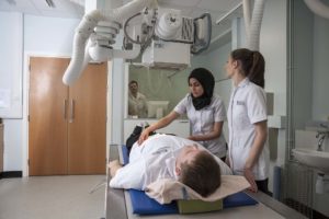
This is a vocational programme with approximately 50:50 ratio theory to practice and is delivered in both the university academic setting and at clinical placement sites throughout the region. The modules, which are delivered at the University, follow four strategic themes. These include: patient centred radiographic practice, anatomy, physiology and pathology, radiation science and research methods. There is an onsite imaging suite and CT scanner to assist in the delivery.
As a student, you will be allocated a hospital placement to attend in several clinical blocks, throughout each of the three years. The focus of each of these placements is closely linked to the academic modules, which are taught using a variety of student centred teaching styles including traditional lectures and small group tutorials. You will also have the opportunity to engage in the award winning team based learning (TBL) approach, an internationally recognised effective teaching method, well evaluated by our current students. You will participate in problem-based learning, where discussions around ‘patient-specific’ scenarios help to enhance your understanding of related issues. You will also be involved in interprofessional learning, which features in all three years of the programme and assists you in understanding the multidisciplinary team (MDT) approach to healthcare.
A continuous clinical assessment scheme, linked to the radiographic practice modules is used in the clinical sites, to record your clinical performance and give you regular feedback, which will enhance your clinical learning. This information is stored online, and is accessible by yourself, clinical placement staff, and academic staff to monitor your progress throughout the programme. During the programme, you will also have the opportunity to enrich your clinical experience by undertaking an elective placement in an imaging department of your choice, which can be locally, nationally or internationally.

We’ve been awarded a Gold rating for educational excellence.
Discover what you'll learn, what you'll study, and how you'll be taught and assessed.
Year one will equip you with foundational knowledge and skills, which will be developed in the subsequent years of the programme. The modules in this year follow the previously mentioned themes: patient centred radiographic practice, anatomy, physiology and pathology, radiation science and research methods.
All modules are compulsory and must be successfully completed before progression to the next year of study.
Programme details and modules listed are illustrative only and subject to change.
The aim of year two is to consolidate the learning experiences from year one and extend them further to provide a foundation for more complex examinations involving specialist equipment. Professional practice will inspire students to become increasingly autonomous, encouraging an appreciation of the challenging issues relating to healthcare.
All modules are compulsory and must be successfully completed before progression to the next year of study.
Programme details and modules listed are illustrative only and subject to change.
The aim of year three is to expand your knowledge of the specialist clinical areas and to promote a level of independence and professional responsibility in preparation for graduation and registration with the Health and Care Profession Council (HCPC). As a qualified diagnostic radiographer you can become a member of the Society of Radiographers.
All modules are compulsory and must be successfully completed before progression to the next year of study.
Programme details and modules listed are illustrative only and subject to change.
Learning is promoted through a wide variety of activities which that enables students to become autonomous and independent continuous learners. An award winning team based learning approach features in many of the modules, along with interactive lectures and student led seminars. Problem-based learning is used to cover patient centred scenarios and collaborative projects are often used to teach research and evidence based practice. The programme has the benefit of an onsite digital imaging suite and CT scanner to enhance clinical skills teaching and there is access to the Human Anatomy Resource Centre, which complements students’ learning.
Throughout the programme there are shared lectures, and tutorials with students from other directorates within the School of Allied Health Professions and Nursing. This is to promote inter-professional education and learning opportunities across all healthcare professions.
Using a mixture of coursework and examination, a range of assessment methods can be seen across the programmes. These include seen and unseen written examinations, essay assignments with specific word lengths, multiple choice questions, case study presentations, video analysis and interactive practical examinations. Assessment of the work-based learning element of all programmes is an important aspect. You will be required to communicate your views orally and in written form; analyse, implement and evaluate your practice; and to extend the research and evidence base of your chosen profession.
The various methods of assessments have been chosen to provide a balance that will permit the undergraduates to demonstrate their intellectual abilities in all areas to the full.
We have a distinctive approach to education, the Liverpool Curriculum Framework, which focuses on research-connected teaching, active learning, and authentic assessment to ensure our students graduate as digitally fluent and confident global citizens.
The Liverpool Curriculum framework sets out our distinctive approach to education. Our teaching staff support our students to develop academic knowledge, skills, and understanding alongside our graduate attributes:
Our curriculum is characterised by the three Liverpool Hallmarks:
All this is underpinned by our core value of inclusivity and commitment to providing a curriculum that is accessible to all students.
Studying with us means you can tailor your degree to suit you. Here's what is available on this course.
University of Liverpool students can choose from an exciting range of study placements at partner universities worldwide.
Spend a summer abroad on a study placement or research project at one of our worldwide partner institutions.
Every student at The University of Liverpool can study a language as part of, or alongside their degree. You can choose:
Diagnostic Radiography students at the University of Liverpool benefit from our experience in delivering more than 100 years of teaching across practical and professionally focused programmes.
Our curriculum is developed and assessed by leading healthcare providers throughout the North West. Many such partners across the North West provide exciting placement opportunities which allows you to bring your studies to life by gaining a breadth of patient-focused practical experience in a region with a particularly diverse population, providing an invaluable insight to future roles.
We place an emphasis on interprofessional learning modules in order to reflect the multi-professional environments you will encounter in today’s healthcare settings.
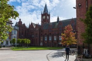
From arrival to alumni, we’re with you all the way:
One of the things I find most interesting about the course is the placement, which offers new experiences every day. When you go into the hospital, you never quite know what you are going to come across. You meet new people and new challenges, developing your technique and communication skills.

Want to find out more about student life?
Chat with our student ambassadors and ask any questions you have.
As a graduate of the School of Allied Health Professions and Nursing, you’ll be eligible to apply for registration with the Health and Care Professions Council (HCPC). You will have gained a qualification that meets the Government’s criteria for ‘fitness for purpose’ and ‘fitness for practice’ as well as developing transferable skills such as communication, information technology, problem solving and teamwork.
You can look to explore careers in:
99% of the School of Allied Health Professions and Nursing students find their main activity after graduation meaningful.
(Graduate Outcomes, 2018-19.)
My qualifications are from United Kingdom.
Your tuition fees, funding your studies, and other costs to consider.
Full-time place, per year - £9,790
Year in industry fee - £1,955
Year abroad fee - £1,465 (applies to year in China)
Full-time place, per year - £32,000
Year in industry fee - £1,955
Year abroad fee - £16,000 (applies to year in China)
The fees shown are for the academic year 2026/27. Please be advised that tuition fees may increase each year for both UK and international students. For UK students, this will be subject to the government’s regulated fee limits.
Tuition fees cover the cost of your teaching and assessment, operating facilities such as libraries, IT equipment, and access to academic and personal support. Learn more about paying for your studies.
We understand that budgeting for your time at university is important, and we want to make sure you understand any course-related costs that are not covered by your tuition fee. This includes costs for specialist equipment, travel to placements, and professional association fees. At the end of year two, students can undertake a self-funded elective placement in the UK or overseas.
Students should expect to cover the following costs:
Home students are able to apply for reimbursement of travel/accommodation costs for placements from the NHS Business Services Authority.
We’re showing the scholarships available to students from United Kingdom.
We offer a range of scholarships and bursaries that could help pay your tuition and living expenses.
If you’re a UK student joining an undergraduate degree and have a household income below £35,000, you could be eligible for a Liverpool Bursary worth up to £2,000 for each year of undergraduate study.
If you’ve spent 13 or more weeks in Local Authority care since age 14, you could be eligible for a bursary of £3,000 per year of study. You’ll need to be a UK student joining an eligible undergraduate degree and be aged 28 or above on 1 September in the year you start.
Are you a UK student with a Black African or Caribbean heritage and a household income of £25,000 or less? You could be eligible to apply for a Cowrie Foundation Scholarship worth up to £8,000 for each year of undergraduate study.
If you’re a UK student identified as estranged by Student Finance England (or the equivalent UK funding body), you could be eligible for a bursary of £1,000 for each year of undergraduate study.
Joining a School of Biosciences degree and have a household income of less than £25,000? If you’re a UK student, you could apply to receive £4,500 per year for three years of your undergraduate course.
Do you live in the Liverpool City Region with a household income of £25,000 or less? Did neither of your parents attend University? You could be eligible to apply for a Nolan Scholarship worth £5,000 per year for three years of undergraduate study.
Are you a UK student with a household income of £25,000 or less? If you’ve participated in an eligible outreach programme, you could be eligible to apply for a Rigby Enterprise Award worth £5,000 per year for three years of your undergraduate degree.
Are you a UK student with a household income of £25,000 or less? Did neither of your parents attend University? You could be eligible to apply for a ROLABOTIC Scholarship worth £4,500 for each year of your undergraduate degree.
Apply to receive tailored training support to enhance your sporting performance. Our athlete support package includes a range of benefits, from bespoke strength and conditioning training to physiotherapy sessions and one-to-one nutritional advice.
Joining a degree in the School of Electrical Engineering, Electronics and Computer Science? If you’re a UK student with household income below £25,000, you could be eligible to apply for £5,000 a year for three years of study. Two awards will be available per academic year.
If you’re a young adult and a registered carer in the UK, you might be eligible for a £1,000 bursary for each year of study. You’ll need to be aged 18-25 on 1 September in the year you start your undergraduate degree.
My qualifications are from United Kingdom.
The qualifications and exam results you'll need to apply for this course.
NHS Values will be assessed in all areas of an application including UCAS Personal Statement and at interview. For more details, please download our explanation of Value Based Recruitment.
| Qualification | Details |
|---|---|
| A levels |
BBB with at least one of the following Science subjects: Biology, Physics, Chemistry or Mathematics.You may automatically qualify for reduced entry requirements through our contextual offers scheme. Based on your personal circumstances, you may automatically qualify for up to a two-grade reduction in the entry requirements needed for this course. When you apply, we consider a range of factors – such as where you live – to assess if you’re eligible for a grade reduction. You don’t have to make an application for a grade reduction – we’ll do all the work. Find out more about how we make reduced grade offers. If you don't meet the entry requirements, you may be able to complete a foundation year which would allow you to progress to this course. Available foundation years: |
| T levels |
T levels in Health, Health Sciences and Science is accepted with an overall grade of Distinction to include in the core. Applicants should contact us by completing the enquiry form on our website to discuss specific requirements in the core components and the occupational specialism. |
| GCSE |
5 GCSEs which must include English Language, Maths and a Science at grades 5 -9 (or grades A* - C if assigned according to previous grading format; Maths and science must be at grade B). Please note that Science dual award is acceptable. Core Science and Applied GCSEs are also considered. All GCSEs should be obtained in one sitting. |
| BTEC Level 3 National Extended Diploma | BTEC Nationals are considered in addition to 5 GCSEs grades 5-9 (or former A* – C), which must include English Language, Maths and a Science. Science dual award, Core Science and Applied GCSEs will also be considered. BTEC National Extended Certificate We will accept one BTEC National Extended Certificate at a minimum of Distinction. This must be accompanied by two A2 subjects at Grade B, of which one subject should include Biology/Human Biology, Physics, Maths or Chemistry. Three separate subjects must be taken between the two qualifications. BTEC National Diploma We will accept in Health and Social Care or Applied Science/ Medical Science graded at DD. This must be accompanied by a Science A2 subject (biology, physics, chemistry or maths) at grade B. In total, between the two qualifications, two separate subjects must be taken. BTEC National Extended Diploma We will accept in either Applied Science/ Medical Science or Health and Social Care at DDD. |
| International Baccalaureate | 30 points to include three higher level (HL) subjects at a minimum of Grade 5. HL Biology must be offered at a minimum of a Grade 6. |
| European Baccalaureate | 74% overall with a minimum mark of 8 in Biology and no other subject less than a 6. |
| Irish Leaving Certificate | Leaving Certificate: 6 Higher Level subjects. 1 subject at grade H1 to include a science subject such as Maths, Physics, Biology or Chemistry, and 2 subjects at grade H2 or above to include a further science subject and or Maths. The remaining 3 subjects must be graded at H3 or above. Out of the six subjects, English, Mathematics and a Science subject must be included. Higher grades may be required from students resitting. |
| Scottish Higher/Advanced Higher | Scottish Certificate of Education |
| Welsh Baccalaureate Advanced | Accepted at Grade B alongside two A2 levels at Grade B, one should be in a Science subject. |
| Cambridge Pre-U Diploma | Will be considered |
| AQA Baccalaureate | Will be considered |
| Graduate application | We welcome applications from graduates holding a minimum of a 2:2 classification. If your degree is not in a Science related subject or it is 5 years or more since you last studied please contact the admission unit for further information. |
| Access | Essential: 60 credits at Level 3, including 15 in credits in biology, 15 credits in maths and 15 credits in physics/chemistry. 39 of the 60 credits must be at distinction, the remaining credits may be gained from ungraded level 3 credits and passed at merit or higher. 3 GCSEs in English, Maths and a science at grades 5/C. |
| Academic Reference | An academic reference must be included within the UCAS application. If the applicant is a graduate and has been working since graduating (within three years), an employer reference is acceptable. |
| Profession-specific knowledge and skills required | The UCAS Personal Statement, must demonstrate understanding of the Diagnostic Radiography role. Applicants should also consider visiting a Therapeutic Radiography department to give them an awareness of the differences between the Diagnostic and Therapeutic Radiography professions. Applicants should have an appreciation of the demands of the programme and a realistic understanding of what is required when on clinical placement. Having experience of working with the general public, children, the elderly or people with disabilities, in a paid or voluntary capacity will strengthen an application. |
| Declaration of criminal background | You will understand that as an Allied Health Professions and Nursing student, and when you qualify, you will be asked to treat children and other vulnerable people. We therefore need information about any criminal offences of which you may have been convicted, or with which you have been charged. The information you provide may later be checked with the police. If selected for interview you will be provided with the appropriate form to complete. |
| Health screening | The University and the School of Allied Health Professions and Nursing has an obligation to undertake health screening of all prospective healthcare students. Any offer of a place on this course of study is conditional on completion of a health questionnaire, and a satisfactory assessment of fitness to train from the University’s Occupational Health Service. This will include some obligatory immunisations and blood tests. The link below provides further information: |
| Disability information | Should a candidate have, or suspect they may have dyslexia, or a long term health condition or impairment that may have the potential to impact upon studies and/or Fitness to Practice, please complete the Disability form. Candidates will then be contacted to discuss requirements for support. |
| International qualifications |
The IELTS requirement is an overall score of 7.0 with no component less than 6.5. Please note – whilst we do accept IELTS qualifications, we do not accept IELTS qualifications that have been sat and gained online. We only accept qualifications that have been sat and gained in person. |
You'll need to demonstrate competence in the use of English language, unless you’re from a majority English speaking country.
We accept a variety of international language tests and country-specific qualifications.
| Qualification | Details |
|---|---|
| IELTS | IELTS 7.0 overall, with no component below 6.5 |
| International Baccalaureate English A: Literature or Language & Literature | Grade 6 at Standard Level or grade 6 at Higher Level |
| International Baccalaureate English B | Grade 7 at Higher Level |
Have a question about this course or studying with us? Our dedicated enquiries team can help.
Last updated 5 February 2026 / / Programme terms and conditions