Environmental sciences
In environmental sciences, we address global challenges by investigating a range of materials from the composition of airborne pollutants, to the micro-structure of rocks for volcanology and earthquake analysis. Using electron microscopy, we can pinpoint sources of contamination, analyse the impact of climate change, and develop effective strategies for environmental protection and sustainability.
Dumortierite Mineralisation Mechanisms
A collaboration between University of Liverpool researchers and The Visvesvaraya National Institute of Technology, India, presents the first comprehensive Electron Backscatter Diffraction (EBSD) study to investigate the mineralisation mechanisms of dumortierite, a relatively rare aluminous borosilicate mineral. The research focuses on vein mineralisation found in metapelitic schists from the Amgaon Gneiss Supracrustals in Central India. Boron-bearing minerals like dumortierite are of high interest for their applications in renewable energy (wind turbine blades), borosilicate glass manufacturing, and health products, despite facing a future risk of supply shortage. Electron microscopy played a pivotal role in the characterisation by using EBSD to reveal that dumortierite needles crystallise along muscovite cleavage surfaces. These advanced microstructural analyses demonstrated that the migration of hydrothermal fluids is facilitated by ripplocations, newly discovered crystal defects, which focus the mineral distribution within muscovite-rich rocks.
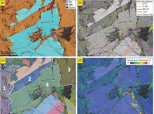
Figure: (a) Phase map of EBSD 1area in Figure 3b. The key shows that red=dumortierite; orange=muscovite; cyan=kyanite. Small yellow arrows point to rounded and serrated Ky phase boundaries; (b) Band contrast image where dark grey indicates low-Kikuchi pattern quality and light grey high Kikuchi pattern quality. The key shows that grain and subgrain boundaries, >15° and <15°, respectively, are colour coded by disorientation angle. (c) All Euler (AE) angle map highlighting in white grains 1, 2, 3 (Ms) and4 (Ky) analyzed using pole figure sin Figure 7. Each colour represents the absolute orientation of a given grain in relation to the SEM reference frame. (d) Kernel average misorientation (KAM) map of the same area. Yellow traces represent low-angle boundary traces with ∼2.5° disorientation across. Topaz (Tpz, colored grey) is labelled and indicated by yellow arrows.
S. Dandekar, E. Mariani, T. R. Dandekar, R. K. Khatirkar, K. Pande, J. Gardner, H. Bagshaw, K. Randive, D. Peshwe. American Mineralogist (2025). https://doi.org/10.2138/am-2024-9380
Deformed Eclogite Rheology
This study by Dave McNamara (et al) examines omphacite-rich eclogites sourced from the Zermatt-Saas unit in the Italian Alps to understand the rheological properties and flow of subducted oceanic crust. Electron microscopy identified a lack of internal strain structures like subgrains, alongside the presence of sharp, asymmetrical chemical zonation within the mineral grains. This was essential in proving that diffusion creep, likely assisted by fluids, acts as the primary mechanism responsible for the rock's lattice preferred orientation (LPO) and overall strength during exhumation. By integrating these microstructural observations with thermodynamic pseudosection modelling, the study demonstrates that diffusion creep can dominate eclogite deformation at high pressure-temperature conditions, challenging previous assumptions that dislocation creep is the sole driver.
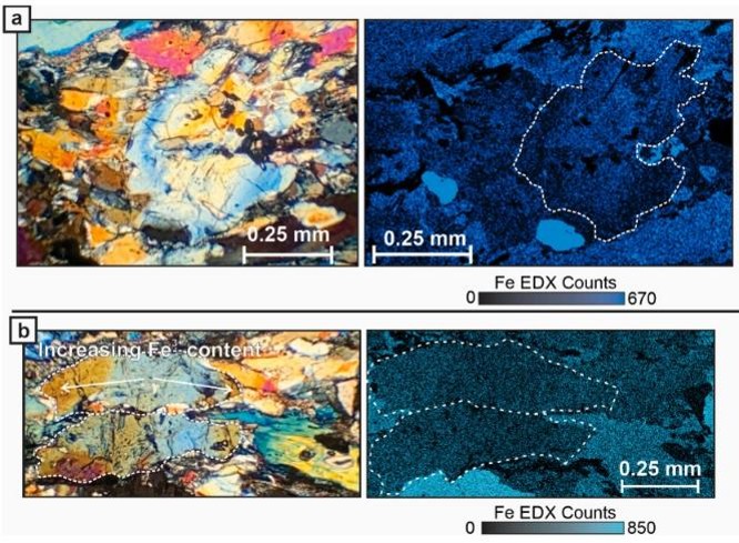
Figure: a) Left: Crossed polarised photomicrograph of a clinozoisite grain that postdates eclogite foliation development from Sample S6.14 showing variable birefringence colours; Right: An Fe EDS map of the same grain showing concentric Fe zoning matching the birefringence patterns. b) Left: Crossed polarised photomicrograph of two foliated clinozoisite grains in Sample S6.14 showing asymmetrical changes in birefringence colours; Right: An Fe EDX map of the same two clinozoisite grains showing weak Fe zoning matching the birefringence pattern. Blue birefringence colours relate to decreased Fe3+ content while orange birefringence colours relate to increased Fe3+ content.
D. D. McNamara, J. Wheeler, M. Pearce, D. J. Prior (2024) Journal of Structural Geology https://doi.org/10.1016/j.jsg.2023.105033