Picosecond view of a martensitic transition and nucleation in the shape memory alloy
RUEDI is equipped with an MeV electron beam to investigate a specimen in terms of electron diffraction and imaging. This allows observations of a specimen with heavier elements as in alloys, in both real and reciprocal spaces.
A shape memory alloy, e.g. Heusler alloy, has caught attention as a functional material in applications for automotive, aerospace, robotic, microelelectromechanical systems and in medical and dental systems.
Although their structure and properties are closely related, it is difficult to fundamentally understand the phase transition, for example, due to the lack of extremely fast probes both in real and reciprocal spaces. The phase transition can happen locally at nanometer scale. As a specimen is not always homogeneous, it is difficult to predict where and when it happens. In addition, a phase transition can happen at a time scale faster than 30psec that is beyond a time scale of synchrotron.
Reference: PHYSICAL REVIEW B 96, 174203 (2017): M Zhang, Gaolong Cao, Huanfang Tian, Shuaishuai Sun, Zhongwen Li, Xingyuan Li, Cong Guo, Zian Li, Huaixin Yang, and Jianqi Li.
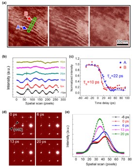
Image: FIG. 2. Time-resolved experimental data for the photoinduced phase transition from the MT phase to AUS phase. (a) Three typical 4D-TEM images obtained at time delays of t = −20, 0, and 20 ps, with a laser-pumping fluence of 5mJ cm−2. (b) Time-dependent profiles for the MT domains in the area outlined by the green rectangle in (a); the ultrafast annihilation of the MT domains is demonstrated. The short lines on each peak show the visible movements of domain walls through the MT transition. (c) Domain intensities obtained from areas A and B as a function of the time delay. The difference in their time constants is attributed to the thickness distinction between area A (50 nm) and area B (30 nm). (d) SAED patterns taken along the [001] zone axis at different time delays; the arrows show visible spot splitting. (e) Ultrafast changes of the spot splitting indicated by the arrows in (d), obtained with a laser-pumping fluence of 10 mJ cm−2, which demonstrate a picosecond transition from the orthorhombic MT phase to the cubic AUS phase. Spot splitting becomes almost invisible for t > 13 ps.