Direct detection of strong THz electromagnetic fields
Unlike XFEL and laser imaging, RUEDI uses a pulsed electron beam. The electron beam interacts both with electric and magnetic fields, making it possible to directly observe the ultrafast dynamics of the electromagnetic field in the microscopic region even in a free space.
A relatively weak electromagnetic field can be detected by using a pulsed electron beam at a lower acceleration voltage. RUEDI's high-energy pulsed electron beam is a rare probe for microscopic imaging.
A strong THz electromagnetic field has been applied as a driving pump. It is conceivable that an electric field that can be as high as 100 MV/m is widely demonstrated in order to induce, for example, phase transition, ionization, carrier manipulation.
When a metallic nanowire is illuminated with a powerful femotosecond laser, it can induce a sub-THz electromagnetic field on its surface. The electron beam crosses the immediate vicinity of the wire and is deflected by both an electric and a magnetic field. This deflection can be directly observed in the reciprocal lattice space, and its spatial distribution is expected to be observed by dark field imaging in the real space.
RUEDI has a large space around a specimen both in electron diffraction and electron imaging. It allows various type of illumination setups.
Reference: Direct detection of strong sub-terahertz surface waves generated on a metal wire by high-intensity laser pulses, Shigeki Tokita, Shuji Sakabe, Takeshi Nagashima, Masaki Hashida & Shunsuke, Inoue Scientific Reports 5:8268 (2015).
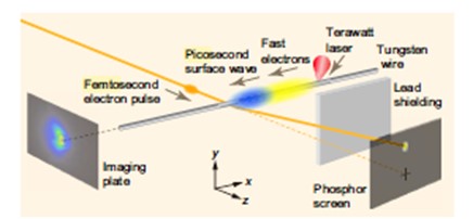
Figure 1: Experimental layout for femtosecond electron deflectometry measurement and emission distribution measurement of fast electrons.
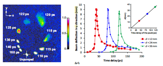
Figure 2: Surface waveform measurement by electron beam deflectometry. (a) Electron beam images detected on the phosphor screen at d 5 36 mm. This is a composite image of 10 single-shot images for different delay durations. (b) Time traces of the beam deflection in the y direction. Inset shows a plot of d versus the time delay of the peak of the time trace. (c) Time traces of the beam deflection in the x direction.