The main experimental techniques we use are photoemission spectroscopy, low energy electron diffraction and scanning tunnelling electron microscopy (explained below). We also use a range of sample handling and preparation techniques such as ultra-high vacuum, in-situ sample changes, heating (1500K), cooling (140K), thin film deposition, and in-situ electrochemistry.
Photoemission
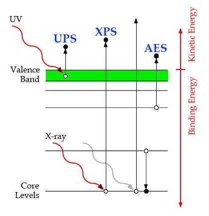
Photoemission spectroscopy (in particular XPS and UPS) determine the binding energies of electrons.
XPS uses x-rays which are able to excite the core electrons, making it very useful for compositional analysis. UPS uses ultra-violet light and excites electrons in the valence band providing information about valence energy levels and chemical bonding.
Low Energy Electron Diffraction
Low-energy electron diffraction (LEED) is used to determine the surface structure of crystals.
By bomarding a surface with a beam of low-energy electrons a diffraction pattern can be observed on a surface. We use a high-resolution video camera to work out how the intensities of diffraction spots change as a function of beam energy (this is called LEED-IV). The LEED-IV curve can then be compared to a theoretical curve to determine information about the position of atoms on a surface.
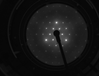
Root 3 structure of I on Au(111)
Scanning Tunneling Microscopy
A Scanning tunneling microscope (STM) is capable of imaging surfaces at the atomic level.
This works by bringing a conducting tip very near to the surface and creating a difference in voltage between the surface and tip, which allows electrons to tunnel through the vacuum between them. The tunnelling current will then depend on the tip position, voltage and local density of states. The tip is scanned across the surface in order to build up a picture of how atoms are arranged on a surface.
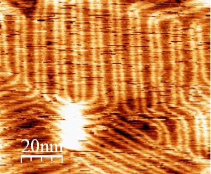
STM image of Au(111) showing the herringbow reconstruction
Equipment
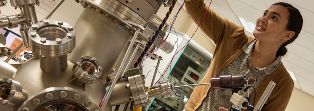
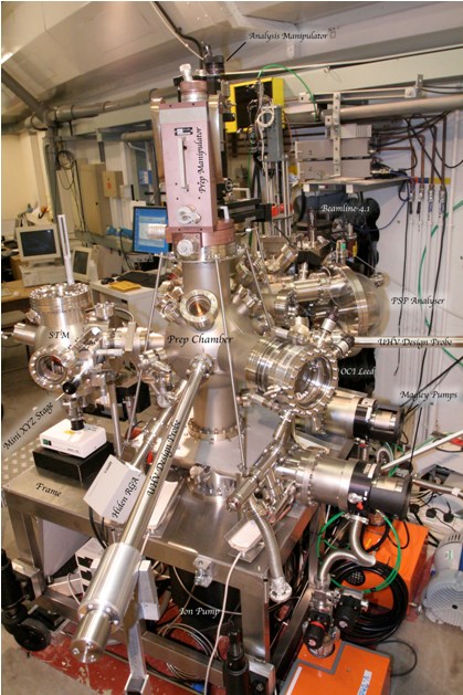
The station shown in the photograph is our primary system. It essentially consists of three spherical chambers mounted on a heavy rigid frame designed to minimise vibrations.
The frame is purposely built to be almost twice as heavy and rigid as those normally used at the SRS (where it was first commissioned). The smaller of the three vessels is a stainless steel chamber housing the Omicron STM-1. The other two chambers are mu-metal vessels, one for analysis comprising a 5-channeltron PSP Vacuum Technology electron energy analyser, a dual anode X-ray source, a UV lamp and standard manipulator. The second mu-metal chamber serves as a preparation chamber and houses a OCI low current MCP based LEED system for I-V measurements, ion gun, a Hiden RGA mass spectrometer and a VG manipulator.
Each of the three UHV chambers are connected via VAT gate valves. Samples can be introduced via a load lock, first into the preparation chamber. Two UHV Design magnetic probes are used to transport samples either into the analysis chamber or onto a micro-XYZ stage before the STM. From the micro manipulator, the sample can be picked up by the STM wobble stick and moved onto the STM stage for imaging.
The sample transfer stage and sample stages on the manipulators in the analysis and preparation chambers are special from Omicron. The sample stages allow heating to 1500 K by indirect irradiative heating. Semiconducting samples such as silicon can be directly heated by the specially designed stages. There is also an e-beam heating stage which is housed in the analysis chamber. By having all three systems of heating, it will be possible to handle sample preparation of almost all types.
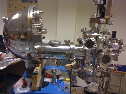
The lab houses a second single chamber UHV system (see picture below), this system consists of a Scienta SES200 electron energy analyser for XPS/UPS measurments. There is also a LEED optics for LEED-IV measurments, and samples can be annealed upto 1300K and cooled to about 140K using standard omicron sample plates.