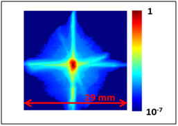
High Dynamic Range Beam Halo Monitor
Halos induced in intense beams by perturbations and other means can lead to beam losses, cause interactions that are detrimental to the efficient transport of the beam and produce high radiation backgrounds. However, measurement of halo is difficult since its intensity can be many orders of magnitude, typically ~10(-5) less than the peak intensity of the beam. We are developing a novel technique to image halo that is adaptive, easy to apply and can be used with any beam radiation source. It employs a digital micro-mirror device (DMD) to generate intensity-based optical masks. Our goal is to achieve a dynamic range (maximum signal to noise ratio) > 10(7). To accomplish this we will employ specially designed filters and masks generated with a DMD to minimize the effects of diffraction and light scattering. An important spin off of this technology is that it can be used to test the performance of any high dynamic range imaging system, e.g. ones that are being developed in astronomy to search for exoplanets.
Optical phase space mapping (OPSM)
We are also developing a DMD based method to perform OPSM, the optical equivalent of the slit-collimator technique commonly used to segment and map the transverse phase space of a charged particle beam. OPSM dispenses with the need to collimate the beam resulting in a great reduction in experimental complexity and cost. With a DMD the process can be readily automated and integrated into an accelerator control system
A key issue is the size and shape of the mask, which must be optimally chosen to minimize diffraction but still maintain a useful resolution and reasonable data acquisition time. To address this problem we are designing and testing different types of mask configurations that can be readily programmed onto the DMD.
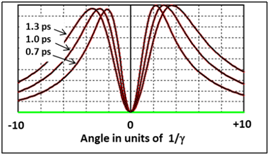
Novel Single Shot, Non-invasive Bunch Length Monitor
This project aims to develop a novel, simple, low cost, non-interceptive, single shot method to measure the bunch length of charged particles generated in an accelerator, a key parameter needed to optimize the accelerator performance. Our technique uniquely employs the angular distribution (AD) of coherent diffraction radiation that is produced as the bunch passes through a simple aperture, e.g. a slit. We have already validated this concept in a proof of principle, time integrated experiment.
Our goal now is to image the entire AD, which will allow bunch length measurement of a single pulse. To accomplish this we are performing experiments at the ALICE accelerator at Daresbury and the FACET accelerator at SLAC using advanced tera Hertz imaging techniques.
Optical Diffraction Radiation Imaging
Optical diffraction radiation (ODR) produced by a charged particle passing by an edge or through an aperture, e.g. a slit or an iris, is a promising beam size monitor for very high energy beams, e.g. LHC and the European XFEL, because it is essentially non-invasive and the radiation generated becomes more intense with energy. Previous studies have concentrated on imaging the far field angular distribution of ODR. However, our analysis shows that imaging the source of the ODR, i.e. the aperture itself, is much simpler and less subject to interference from upstream radiation sources. A key issue and limiting factor is the point spread function (PSF) of ODR, which determines the resolution and dynamic range of the measurement. Our goal is to measure and utilize the PSF to determine the beam size from the ODR source field image produced by a high energy beam. For this purpose we will perform experiments at high energy (GeV) electron beam facilities, e.g. the Swiss FEL and the KEK ATF accelerators.
Currently involved Quasars
Selected publications
- H. Zhang, R. Fiorito, A. Shkvarunets, R. Kishek, C. Welsch, Phys. Rev. ST Accel. Beams 15, 072803 (2012)
- R. Fiorito, H. Zhang, A. Shkvarunets, et. al. Proc. of BIW12, Newport News,VA (2012)
- R. Fiorito, A. Shkvarunets and P. O’Shea, AIP Conf. Proc. 648, p.,187-194 (2002)
- B. Riddick, E. Montgomery, R. Fiorito, et. al., Phys. Rev. ST Accel. and Beams, 16, 062802 (2013)
- A. Shkvarunets, R. Fiorito, et. al. Proc. of DIPAC07 (2007)
- R. Fiorito and D. Rule, Nucl. Instr. and Meth. B 173, 67 (2001)
- P. Karataev, et. al., Phys. Rev. Lett. 93, 244802 (2004)
- R. Fiorito, D. Rule and W. Kimura, AIP Conf. Prod. 472, p.725 (1999)
- D. Xiang, et. al. Phys. Rev. ST Accel. and Beams, 10, 062801 (2007)
- T. Aumeyr, et. al. Phys. Rev. ST Accel. Beams 18, , 042801 (2015).
Back to: QUASAR Group