These systems are three independent “cubes”. The β-cube is a high sensitivity PET system with a sub-millimetre resolution, which can provide PET scans of the whole body of a mouse in a single imaging frame and rats in a number of frames.
The ɣ-cube is a high resolution, high sensitivity collimator based SPECT system.
Finally the x-cube is a high throughput µCT scanner which is used to acquire anatomical images to co-register with PET and SPECT data.
All cubes are compatible with the same heated animal beds, which monitor the animal’s vital signs, and ensure the animals remain in a consistent position during imaging in each of the cubes enabling easy and accurate co-registration of the images.
Below are examples of images and videos taken using the systems.
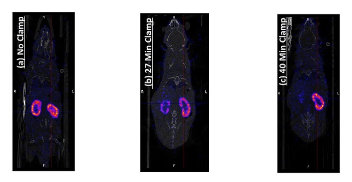
- Assessing kidney function
Kidney function assessed using 99mTc dimercaptosuccinic acid (DMSA) - single photon emission computerized tomography (SPECT) imaging. The right kidney underwent ischemia reperfusion injury. (a) before clamping, (b & c) reduced perfusion in the injured kidney post clamping.
- Dynamic Positron Emission Tomography (PET) image overlaid on a Computed Tomography (CT) image
(a) Uptake and clearance of radio tracer
Uptake of 18F-fluorodeoxyglucose (18F-FDG) initially in the lungs and later into the kidneys and clearance to the bladder.
(b) Imaging Tumor Glycolysis
High uptake of 18F-FDG seen in the F98 glioma implanted subcutaneously (shown by white arrow)
Currently active projects:
- Measurement of tumour progression
Back to: Research