It enables high sensitivity, non-invasive, imaging permitting monitoring of processes such as disease progression, cell trafficking, and gene expression patterns in living animals.
Below are examples of images and video taken using the system.
- Cell bio-distribution - Scarfe et al. Stem Cell Research & Therapy 9:332 (2018)
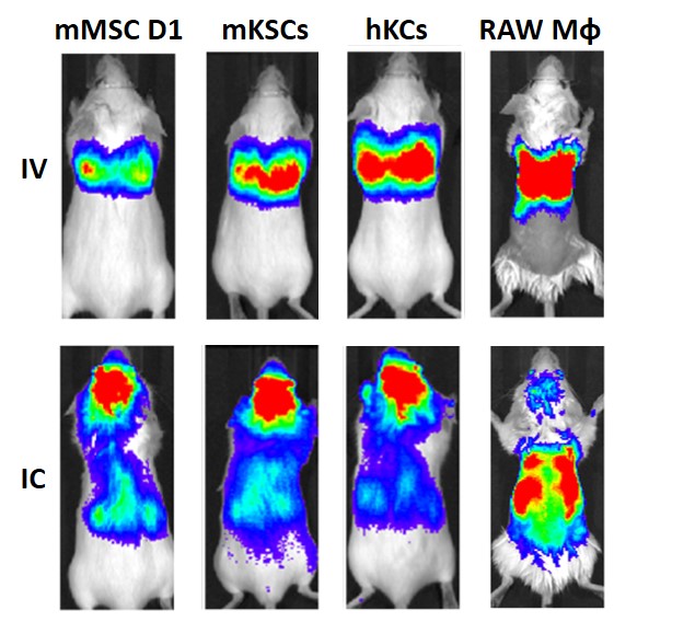
Biodistribution of different cells following intravenous (IV) or intracardiac (IC) administration. BLI immediately after administration, showing that cells were always confined within the lungs after IV administration, but distributed throughout the body after IC administration.
- Longitudinal tumour monitoring - Scarfe et al. Stem Cell Research & Therapy 9:332 (2018)
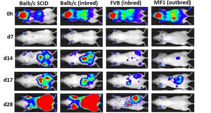
Assessing the safety of cell transplantation using BLI.
Transplanted cells form teratomas (tumours) in two-three weeks after injection regardless of the mouse strain used indicating the potential risk with the use of mesenchymal stem cells.
- Monitoring tumour burden in vivo (chick model)
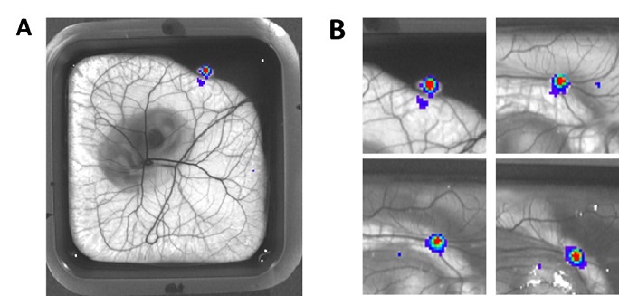
Mesothelioma cell line (MESO-8T) stably expressing luciferase was implanted on the chorioallantoic membrane (CAM) at embryonic day 7 (E7). Following administration of luciferin, bioluminescent signal can be seen in the resulting tumour nodules (A). Bioluminescent imaging was performed on different days (B) to quantify and monitor tumour growth.
Back to: Research