It is specifically geared for small animal imaging and spectroscopy studies as well for high-resolution imaging of ex vivo and non-biological samples. Besides 1H MRI and MRS, the scanner can also be used for observing other nuclei such as 13C, 31P, 23Na, and 19F.
Below are examples of videos and images taken using the system.
- Automated segmentation of magnetic resonance images for mouse brain phenotyping
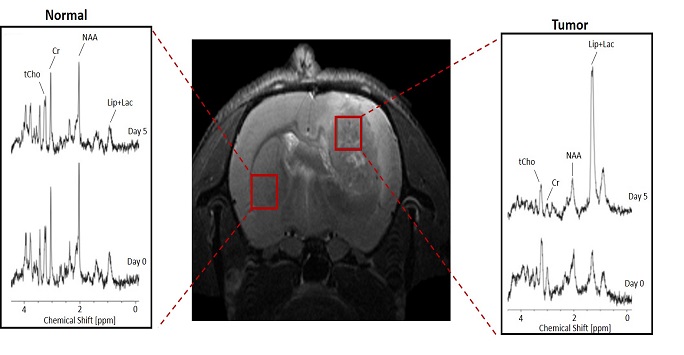
- Magnetic Resonance Spectroscopy (MRS) showing the difference between normal brain and tumor in a rat model
- Magnetic Resonance Imaging (MRI) of an ex vivo mouse kidney
- Short-axis view of the motion of a mouse heart over one cardiac cycle
Currently active projects:
- High resolution MR microscopy for MR phenotyping of genetic models;
- In vivo cell tracking for assessing the safety and efficacy of cell therapies;
- Non-invasive measurement of blood flow in kidneys, brain and tumours;
- Measuring tissue metabolism in vivo (MR spectroscopy, chemical shift imaging) in liver and brain tumours;
- Diffusion tensor imaging (DTI) in brain tumours and tissue phenotyping;
- Cardiac imaging;
- MR relaxometry using T1, T2 mapping for contrast agent characterization.
Back to: Research