Two camera-based spinning disk confocal imaging systems providing optical sectioning and high image capture rates.
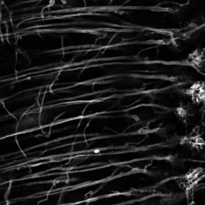
Axons (long “tubes”) and synaptic terminals (right) in the Drosophila fly visual system labelled with a plasma membrane marker. Data courtesy of the Sanchez-Soriano lab.
Microscopes:
- LSM 900 (Nuffield building)
- LSM 900 (William Henry Duncan building)
- LSM 880 MP
- LSM 880 BioAFM
- LSM 800
- LSM 780
- Leica SP5
- 3i Marianas spinning disk
- Andor Dragonfly spinning disk
Back to: Research