Combining these methods allows to image across scales and probe different parameters like topography or surface stiffness.
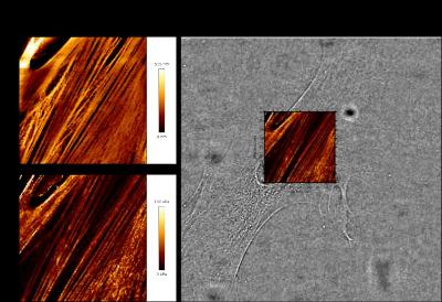
The above cells from a patient suffering from Hutchinson-Gilford Progeria Syndrome were imaged on the microscope, both in atomic force microscope and light microscope mode. The cells were imaged whilst alive. In AFM mode, the topography of the cell was recorded, resulting in a height map. Simultaneously, the stiffness of the same region was also recorded and is displayed as a stiffness map. Using an integrated camera, a transmitted light image of the cell was captured and the stiffness map has been overlaid for reference. Data courtesy of Tom Waring (Zech lab).
Microscope:
Back to: Research