In a fight between detailed or simple models, simplicity wins… for now.
Thomas Primidis and his collaborators Dr. Vadim Soloviev from Adaptix Ltd and Prof. Carsten Welsch from the Quasar Group have published their recent research outcomes in IOP's Biomedical Physics and Engineering Express (https://doi.org/10.1088/2057-1976/ac1987). They have shown that when simulating a digital tomosynthesis procedure, conventional Monte Carlo models are accurate in simulating radiation transport inside human tissues and that using more realistic but also hard to develop models does not significantly change the simulation outcome. Conventional models consider human tissues to be composed of independent atoms as if they were all in gaseous form. This makes for a convenient theoretical description and such a model has been implemented in almost all Monte Carlo codes.
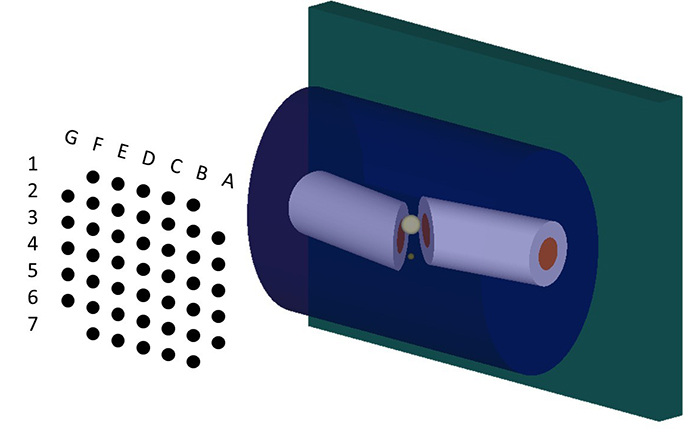
Schematic of a Digital Tomosynthesis simulation using an X-ray source array and a flat panel detector. The phantom resembles a broken limb. (Image credit: Primidis et al 2021 Proc IPAC21 TUPAB409, CC BY 3.0)
However, it is well known that even liquid water has a small-scale molecular order and this is true for human tissues too. The practical effect of this Independent Atom approximation Model (IAM) is that in theory the probability of coherent (Rayleigh) scatter peaks at 0 scattering angle, while in reality non-zero values are observed due to the low range molecular order. This model was found by previous researchers to cause non negligible changes in simulated 2D X-ray images of the human body however when a 3D CT reconstruction was applied, those changes became insignificant. Thomas et al. tested what happens in the case of digital tomosynthesis, a modality that offers 2.5D information, offering a combination of the low dose benefits of 2D radiography and the depth resolution of CT.
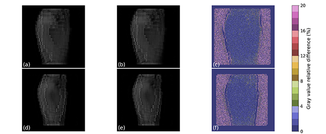
(a), (d) two tomosynthesis slices of the right hand of the ICRP 110 voxel phantom using the IAM model; (b), (e) the same slices after adding molecular interference effects. The differences between the two models are below 4% for both slices as shown in (c) and (f). (Image credit: Thomas G Primidis et al 2021 Biomed. Phys. Eng. Express 7 055016, CC BY 4.0)
The results showed that the digital tomosynthesis reconstruction algorithm washes away those differences like the CT reconstructions do, therefore there is no need for researchers to update their codes with more realistic models.
Digital tomosynthesis has been proposed as a lower dose alternative to CT or as a higher diagnostic quality alternative to bedside 2D radiography. This has resulted in a re-sparked interest and R&D to bring more tomosynthesis systems in clinics. The results by Thomas et al. will be pivotal in accelerating this R&D, especially in deciding the correct tools to do it with.
Thomas Primidis said: “It was very reassuring that these simple models can still do the job accurately. However, the accelerated R&D in novel detectors and X-ray sources might soon allow X-rays, CT and Digital Tomosynthesis scans to diagnose diseases that are expressed on a molecular level. Breast and brain imaging and early diagnosis in general would really benefit from that. When that happens, accurate modelling of molecular effects in radiation transport will be elemental during design and optimisation of the next generation of diagnostic technology."
Further information:
'Accuracy of the independent atom approximation in digital tomosynthesis Monte Carlo simulations'
Thomas G Primidis et al 2021 Biomed. Phys. Eng. Express 7 055016 (2021)
https://doi.org/10.1088/2057-1976/ac1987