CANCELLED Molecular Simulation of Biomolecular Assemblies
1:00pm - 2:00pm /
Monday 9th December 2019
/ Venue: Lecture Theatre 1 Life Sciences Building
- Jill Madine
- Suitable for: Those interested in Genomes, Systems and Therapeutic Targeting
- Admission: Free
- Book now
Add this event to my calendar
Click on "Create a calendar file" and your browser will download a .ics file for this event.
Microsoft Outlook: Download the file, double-click it to open it in Outlook, then click on "Save & Close" to save it to your calendar. If that doesn't work go into Outlook, click on the File tab, then on Open & Export, then Open Calendar. Select your .ics file then click on "Save & Close".
Google Calendar: download the file, then go into your calendar. On the left where it says "Other calendars" click on the arrow icon and then click on Import calendar. Click on Browse and select the .ics file, then click on Import.
Apple Calendar: The file may open automatically with an option to save it to your calendar. If not, download the file, then you can either drag it to Calendar or import the file by going to File >Import > Import and choosing the .ics file.
Speaker: Jamshed Anwar (Lancaster University)
Accessing molecular assemblies of biomolecules is challenging, given that they are soft-matter. The definitive method is X-ray diffraction but this requires crystals – and the molecular packing in crystals may not reflect that in the biological environment. Cryo-electron microscopy is another option but does not yet give atomic resolution. An alternative is to resort to molecular simulation, wherein the molecular trajectories are simulated using Newtonian mechanics driven by inter-molecular forces. Molecular simulations by design converge to lower free energy, akin to real systems.
I will present some recent simulation results on two biological systems: the self-assembly and molecular organization of tight junction proteins, and the interaction of curcumin with amyloid structures.
Tight junctions are complex multiprotein structures found in epithelial and endothelial cells which serve as selective barriers and regulate the diffusion of small molecules and ions through the intercellular space. They are composed of a network of strands that encircle the cells belt-like. The molecular organization of the tight junction strands is not entirely resolved but is essential to understanding normal physiological function as well as dysfunction in pathological states. Furthermore, the development of new therapeutic strategies will rely on a molecular level understanding of the tight junction barrier mechanism. In the simulations we have explored both the cis(same cell membrane)- and trans(adjacent cell membranes)-interaction with a view to understanding how tight junction strands form and their molecular architecture.
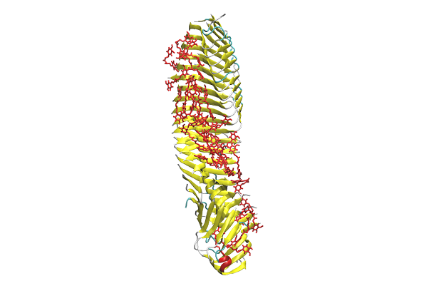
Curcumin is a natural fluorescent dye that binds preferentially to Aβ fibrils. A recent proof-of-concept study has demonstrated its use as a non-invasive Aβ-biomarker for Alzheimer’s disease in live patients, where the curcumin fluorescence was employed to detect amyloid deposits in the retina [1]. We have investigated the molecular basis for curcumin’s specificity for Aβ, simulating the interaction of curcumin with Aβ at multiples levels: with the Aβ monomer, an oligomer, and a pre-formed protofilament
Accessing molecular assemblies of biomolecules is challenging, given that they are soft-matter. The definitive method is X-ray diffraction but this requires crystals – and the molecular packing in crystals may not reflect that in the biological environment. Cryo-electron microscopy is another option but does not yet give atomic resolution. An alternative is to resort to molecular simulation, wherein the molecular trajectories are simulated using Newtonian mechanics driven by inter-molecular forces. Molecular simulations by design converge to lower free energy, akin to real systems.
I will present some recent simulation results on two biological systems: the self-assembly and molecular organization of tight junction proteins, and the interaction of curcumin with amyloid structures.
Tight junctions are complex multiprotein structures found in epithelial and endothelial cells which serve as selective barriers and regulate the diffusion of small molecules and ions through the intercellular space. They are composed of a network of strands that encircle the cells belt-like. The molecular organization of the tight junction strands is not entirely resolved but is essential to understanding normal physiological function as well as dysfunction in pathological states. Furthermore, the development of new therapeutic strategies will rely on a molecular level understanding of the tight junction barrier mechanism. In the simulations we have explored both the cis(same cell membrane)- and trans(adjacent cell membranes)-interaction with a view to understanding how tight junction strands form and their molecular architecture.
Curcumin is a natural fluorescent dye that binds preferentially to Aβ fibrils. A recent proof-of-concept study has demonstrated its use as a non-invasive Aβ-biomarker for Alzheimer’s disease in live patients, where the curcumin fluorescence was employed to detect amyloid deposits in the retina [1]. We have investigated the molecular basis for curcumin’s specificity for Aβ, simulating the interaction of curcumin with Aβ at multiples levels: with the Aβ monomer, an oligomer, and a pre-formed protofilament