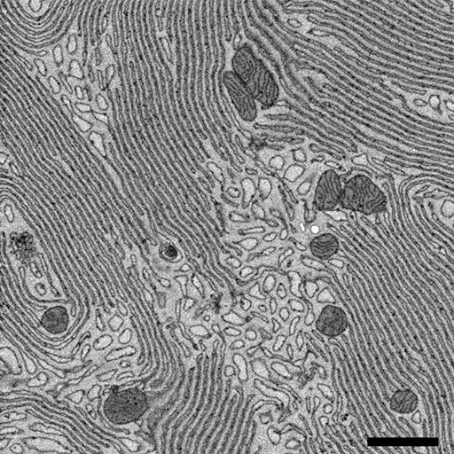
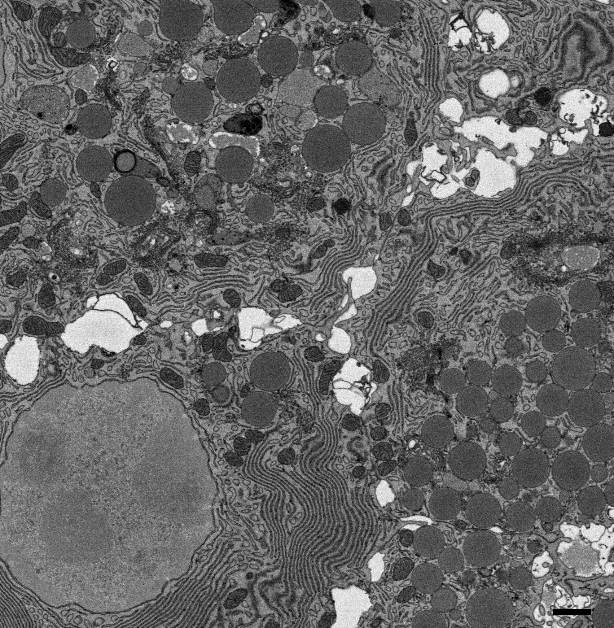
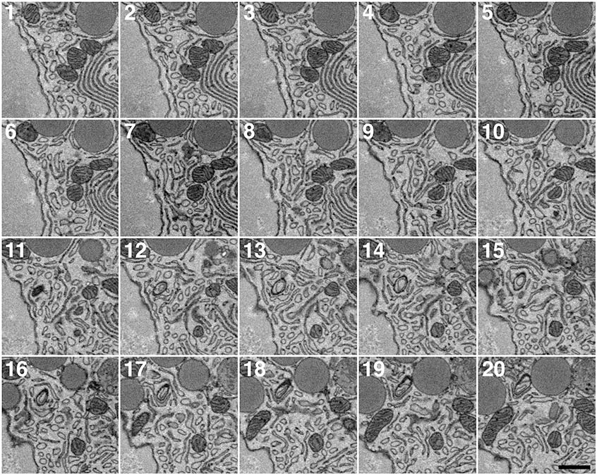
Equipment

The EM Unit contains all of the equipment necessary to analyse biological samples for transmission electron microscopy (TEM). We are also able to offer high-resolution scanning EM (SEM) that includes the ability to look at fully hydrated samples. Using the Gatan 3View system, we can generate serial images through tissue or cell samples for 3D-EM imaging. Over £1 million has been invested in equipment and infrastrucuture since 2004.

Transmission Electron Microscope

In April 2005 we installed an FEI 120kV Tecnai G2 Spirit BioTWIN.

Some of the capabilities of this microscope include:

  • Completely PC driven microscope with individual user defined profiles
  • Digital image capture using SIS Megaview III camera and data analysis with AnalySIS Pro
  • 0.36 nm point resolution
  • Motorised stage
  • Low dose mode to minimise beam damage to sensitive samples
  • Automated controls including alignment and astigmatism correction
  • Cryo-EM capability for frost-free examination of frozen-hydrated suspensions and sections. A Vitrobot is available for highly reproducible cryo-sample preparation
  • Automated electron tomography using 3D Xplore making 3D image modeling possible through a ±70° tilt series.

Various specimen holders are available including the Gatan ±70° cryo-transfer holder and the FEI ±70° single-tilt tomography holder.

Scanning Electron Microscope

In August 2013 we installed an FEI 250 Quanta FEG-ESEM.

Some of the capabilities of this microscope include:

  • Completely PC driven microscope with individual user defined profiles
  • High vacuum (<6x10⁻⁴ Pa) , low vacuum (10-130 Pa) and ESEM (10-4000 Pa) modes
  • BSE and SE detectors for all vacuum modes
  • Resolution ≤1.4nm at 30kV
  • Wet-STEM capability
  • Capability to image fully hydrated samples in ESEM mode.
3View - 3D-EM imaging

The 3View system is a microtome that sits within a scanning EM. A sample embedded in resin is sectioned and then the block face is imaged, this is then repeated to generate a 3D volume of high resolution serial images. 

Download further information (pdf).

Resolution is typically ≥5-10 nm / pixel, sections can be 30-200nm thick, and an image can be taken every minute. This means that from an overnight run 500-1000 images can be taken representing up to 200µm of depth. Typically we are imaging 10-100µm3 volumes overnight to observe anything from organelle organisation through to cell distributions within a tissue or model organism.

Example high resolution images can be seen below. Click on the image for a higher resolution view.

Pancreatic acinar cell
Pancreatic acinar cell. Parallel lines of endoplasmic reticulum (ER) with associated mitochondria. The ribosomes on the ER have not been stained. Bar = 1000nm.
Pancreatic acinar cell. Nucleus, secretory granules, ER, golgi stacks, lysosomes and mitochondria can be seen in a group of 3 cells. Bar = 1000nm.
Montage of 20 high resolution serial sections.
Montage of 20 high resolution serial sections. Nuclear pores in nucleus can be seen on the left. ER and mitochondrial networks extend through the images. Bar = 500nm.
Sample preparation equipment

Our lab is fully equipped for reagent preparation including gold probes and coated grids, and for processing samples through a wide range of EM methodologies.

Equipment specifications:

Specialised equipment includes:

  • RMC EMP 5160 Automated Tissue Processor
  • Emitech K950X Turbo evaporator
  • Quorum Q150T Sputter coater (Au/Pd, Au and Pt targets available) 
  • Quorum K850 critical point dryer
  • Leica EM IGL grid stainer.

Ultramicrotomes:

  • 2x Leica UC6 microtomes with FC6 cryochambers allowing resin or cryo-sectioning
  • Reichert Ultracut S with CFS adaptor for cryosectioning
  • Reichert Ultracut E for resin sectioning.

Cryo-preparation:

  • Leica EMPACT2 high pressure freezer with rapid transfer system enabling reproducible vitreous freezing and correlative light and electron microscopy
  • Vitrobot; automated vitrification system
  • Leica EM MM80 impact freeze
  • Leica AFS2 freeze substitution apparatus with Leica FSP automatic processing system
  • Leica AFS freeze substitution apparatus.