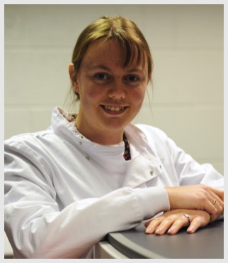
Dr Joscelyn Sarsby
Postdoctoral Researcher
LabA, Centre for Proteome Research
Institute of Integrative Biology
University of Liverpool,
Crown Street
Liverpool L69 7ZB
Tel: +44 151 794 5344
Tel: +44 151 795 4392
Email: jsarsby [at] liverpool.ac.uk
Postdoctoral Researcher
LabA, Centre for Proteome Research
Institute of Integrative Biology
University of Liverpool,
Crown Street
Liverpool L69 7ZB
Tel: +44 151 794 5344
Tel: +44 151 795 4392
Email: jsarsby [at] liverpool.ac.uk

I completed my undergraduate degree in 2010 at the University of Reading reading chemistry with a year in industry, where my final year project was “Matrix-assisted laser desorption ionisation mass spectrometry imaging of Arabidopsis thaliana metabolites.” I used mass spectrometry imaging and cryo-SEM imaging of flowers and siliques of highly unstable glucosinolates to give insight to plant defence mechanisms.
My postgraduate studies took place in a Doctoral Training Centre at the University of Birmingham where I completed a MSc in the Physical Science of Imaging for the Biomedical Sciences in 2011, which saw the integration of chemistry, biology, physics and computer science. The three mini-projects involved included developing a MATLAB code for the semi-automatic reconstruction of phase contrast microscopy images, investigating sample preparation methods for mass spectrometry imaging analysis of whole bovine eyes and the quantification of fluorophores on artificial lipid bilayers.
The PhD aspect of the DTC was completed in two labs at the University of Birmingham under Dr Josephine Bunch and Prof. Helen Cooper, Investigating liquid extraction surface analysis (LESA) and MALDI mass spectrometry imaging of large and small molecules in human liver disease. The aim was to develop analytical methods that would ultimately be used in the study of non-alcoholic liver disease. Initial investigations assed the capabilities of using LESA for imaging and optimising sampling protocol for lipids and proteins. Analysis of the fatty acid binding proteins was explored further, in particular to the identification of a single amino acid substitution that is associated with non-alcoholic steatohepatitis, a potentially fatal liver disease. Both top-down and bottom-up proteomics techniques were employed. Bottom-up resulted in over 300 proteins being detected and identified, including several other known biomarkers for liver disease, however failed to distinguish between two proteins with a single amino acid substation. The top-down approach was successfully employed to identify these two proteins. Field asymmetric ion mobility spectrometry (FAIMS) analysis was coupled with LESA to enhance the quality of intact proteins mass spectra and doubled the number of proteins detected. FAIMS analysis was able to separate lipid and proteins that are extracted simultaneously as well as removing background noise from large unresolvable ions. To fully appreciate the data that was acquired using the FAIMS, visualisations software was developed. In addition to FAIMS, traveling wave ion mobility spectrometry (TWIMS) was also coupled to LESA via the Synapt G2-S. In addition to the separation of lipids from proteins several lipid also showed a separation in drift. MS/MS of one of these lipids revealed that the composition of the two drift peaks differed. The ability to separate isobaric lipids made this technique partially useful for imaging. MALDI imaging was performed with TWIMS, to show the isolation of specific lipid species MS/MS imaging and TWIMS imaging were performed. The MALDI gave a complementary approach to LESA for tissue analysis.
cont’d…
B.Sc. University of Reading
Ph.D. University of Birmingham
Ph.D. University of Birmingham
cont’d…
I am currently funded by the Technology Directorate to run the mass spectrometry imaging instruments in the Centre for Proteome Research, to image metabolites, lipids, and peptides.
Publications
Sarsby, J., et al., Liquid Extraction Surface Analysis Mass Spectrometry Coupled with Field Asymmetric Waveform Ion Mobility Spectrometry for Analysis of Intact Proteins from Biological Substrates. Anal Chem, 2015. 87(13): p. 6794-800.
Sarsby, J., et al., Top-down and bottom-up identification of proteins by liquid extraction surface analysis mass spectrometry of healthy and diseased human liver tissue. J Am Soc Mass Spectrom, 2014. 25(11): p. 1953-61.
Griffiths, R.L., et al., Formal lithium fixation improves direct analysis of lipids in tissue by mass spectrometry. Anal Chem, 2013. 85(15): p. 7146-53.
Sarsby, J., et al., Mass spectrometry imaging of glucosinolates in Arabidopsis flowers and siliques. Phytochemistry, 2012. 77: p. 110-8.
