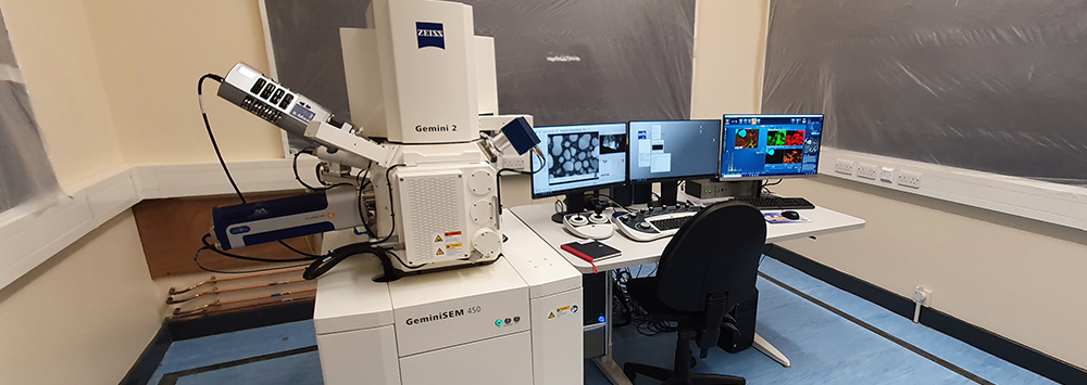
The Zeiss GeminiSEM 450 is a state-of-the-art variable pressure (VP) FEG SEM, fitted with Schottky field emission gun.
Technical specifications
Accelerating voltage: 0.02 kV to 30 kV, continuously variable in 0.01 kV steps.
Beam current: 3 pA to 40 nA with continuous adjustment simultaneous with optimized spot size.
Resolution: 0.7 nm at 15 kV, 1.1 nm at 1 kV; 1.0 nm at 15 kV and 1.4 nm at 3 kV when using VP.
The unique design of the Gemini column means that we can image magnetic samples in high resolution 100% distortion free, as well as tiled samples at high depths of field.
Detectors
- The new high-speed Symmetry EBSD detector, based on CMOS technology (Oxford Instruments)
- X-Max 50 mm2 EDS detector (Oxford Instruments)
- DEBEN panchromatic cathodoluminescence (CL) detector
- Variable pressure secondary electron (VPSE) detector
- In-lens and in-chamber secondary electron (SE) and backscatter electron (BE) detectors, also used in VP mode.
The Zeiss GeminiSEM 450 is optimised for high-resolution, high-speed EBSD data acquisition on samples from nm scale to large areas of several cm2, and also for simultaneous EBSD and EDS. This means that we can determine the full crystallographic orientation and chemistry of minerals and rocks, metals, ceramics and advanced materials. We also offer additional capability for transmission Kikuchi diffraction (TKD) analysis of electron transparent samples.
Post-acquisition softwares include:
- Aztec Crystal: for EBSD post processing and data analysis
- CrossCourt Rapide: for strain analysis by high-angular resolution (HR) EBSD,
- Mountains: for 3D model rendering and quantification of surface topography
In addition, ultra-high-resolution secondary electron images show 3D nanometer scale surface details at voltages as low as 1 kV. With the VP system we can analyse non-conductive and vacuum sensitive materials. The large chamber is designed for use with hot, cryo, deformation and fluid-solid phase in-situ stages.
Back to: Scanning Electron Microscopy Shared Research Facility (SEM SRF)
