Researchers
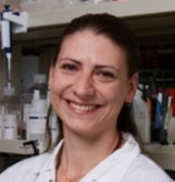
Dr Cordelia Dunai
Postgraduate Research Associate

We are studying mouse and hamster models of SARS-CoV-2 infection to further understand brain complications of COVID-19. We find that even in the absence of direct viral infection, there is immune activation in the brain and we aim to investigate how to prevent and treat brain complications from COVID-19 using this model.
A lot of information can be gathered by measuring proteins in blood-- called biomarkers. Our study measured different types of biomarkers in participants' blood that relate to different health events. Participants who had neurological complications from COVID-19 were found to have higher levels of proteins that are released from damage to the nervous system and these are a useful, measurable biomarker for future therapeutic strategies.
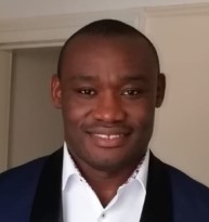
Dr Franklyn Egbe Nkhongo
Postgraduate Research Associate

My main research activity is assessing diagnostics and prognostics biomarkers of neurological insults and immunology/inflammatory mediators in COVID-19 and Encephalitis patients. We use ultra-sensitive bead-based digital multiplex immunoassay to quantify clinical biomarkers in patients' samples to inform on clinical outcomes, likely disease mechanistic, potential therapy targets and for prognostics purposes. The main objective is to better understand and manage neurological complications in COVID-19 and Encephalitis. We use the Quanterix SR-X simoa platform to quantify biomarkers of brain injury, neurofilament light (NF-L), total tau, glial fibrillary acidic protein (GFAP), ubiquitin carboxyl-terminal hydrolase L1 (UCH-L1), phosphorylated Tau (p-Tau-181), Aβ40, and Aβ42 and the Bio-Plex Pro Human 48-Plex Cytokine Panel (Bio-Rad) to quantify 48 immunology biomarkers.
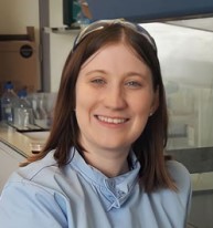
Dr Claire Hetherington
Postgraduate Research Associate

In my postdoctoral research I am investigating the critical connection between COVID-19 infection, the body’s immune response, and damage to the brain. When someone gets infected with COVID-19, their body's immune response is triggered, causing various cells to release signalling molecules called cytokines. The cells that line blood vessels lay a crucial role in maintaining the blood-brain barrier, which is a protective barrier that keeps harmful substances out of the brain. However, when exposed to COVID-19 and cytokines produced by immune cells, endothelial cells can become activated and start producing even more inflammatory cytokines that enter the brain. This, in turn, leads to activation of microglia and astrocytes, which are special cells in the brain responsible for immune responses and supporting neuronal function. However, when these cells are excessively activated due to the cytokine signals, it can result in damage to neurons and, in severe cases, even neuronal death.
To study this, we can grow all these types of cells in the laboratory, find which molecules are produced in response to virus exposure and see how the different types of cells respond to them. This is called “in vitro modelling” as we are modelling what happens in the brain. By doing so, we can determine the pathway of events that lead to neuronal damage. By understanding this pathway, scientists can develop strategies to mitigate the neurological effects of the virus and potentially find new ways to protect the brain during such infections.
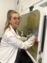
Ms Sarah Boardman
PhD Student

I’m a fourth-year PhD student investigating how Herpes Simplex Virus Type 1 (HSV-1) disrupts the blood-brain barrier (BBB) and contributes to encephalitis. To explore these interactions, I develop biologically relevant in vitro BBB models that help uncover viral mechanisms and identify potential therapeutic interventions.
One of the models I use incorporates microfluidic chip technology—small devices with microchannels cut or moulded into materials such as silicon, glass, or polymers like polydimethylsiloxane (PDMS). These channels are arranged in specific designs for targeted applications and are often interconnected. Microfluidic chips allow for the spatial separation of different cell types, closely mimicking the natural architecture of the BBB. This setup enables endothelial, neural, and glial cells to be cultured under optimal conditions, enhancing the physiological relevance of the model.
In my microfluidic model, I culture a 3D endothelial lumen and use a syringe pump to simulate haemodynamically relevant shear flow. Astrocytes are co-cultured in an adjacent channel, allowing communication through microporous interfaces. To assess barrier integrity, I use transendothelial electrical resistance (TEER) measurements and image tight junction proteins to evaluate their expression levels. Following HSV-1 infection, I assess the impact on barrier function and monitor changes in cytokine expression to understand the immune response.
In addition to the microfluidic model, I have developed a second in vitro system using 96-well TEER/impedance plates compatible with a multielectrode array (MEA) biosystem. This setup allows me to monitor barrier integrity in real-time and has revealed a dose-dependent response in brain endothelial cells following direct or indirect HSV-1 infection. I also examine cytokine expression via ELISA and aim to investigate whether targeted adjunctive therapies can reduce BBB damage caused by HSV-1.
Future work will involve microfluidic chips that integrate with the MEA system and additional BBB cell types to further enhance the complexity and relevance of the models.
Ms Bethany Facer
PhD Student

My research interest is neuroimaging in disease populations. I am working on the DexEnceph study investigating imaging alterations in herpes simplex virus encephalitis and the associations with inflammatory markers. I am also a member of the neuroimaging working group for the COVID Clinical Neuroscience Study (COVID-CNS) where I coordinate and analyse the data. At present I am focusing on a population of people with SARS-CoV-2 induced movement disorders.
Ms Anne Caroline Marcos
PhD Student

I am a Brazilian neuroscientist and third-year PhD candidate at Fiocruz, Brazil, currently undertaking a research placement at the Brain Infections & Inflammation Group (BIIG), University of Liverpool. My research explores how neurotropic viral infections, including Herpes simplex virus type 1 (HSV-1) and Zika virus, impact neuronal health and mitochondrial function. Using human iPSC-derived neurons, I investigate how viral infection alters mitochondrial dynamics, energy production, and stress responses, and how these changes may contribute to neuronal dysfunction and neurodegenerative processes. This work supports the group’s broader aim of understanding how infectious agents interact with the nervous system and affect brain function.