| PLANT TISSUE CULTURE
CASE STUDY 3 Demonstration of tissue culture for teaching |
||||||
| INTRODUCTION WHAT IS IT? USES CASE STUDY 1 Anther culture for cold hardiness CASE STUDY 2 CASE STUDY 3
|
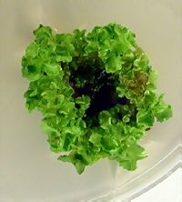
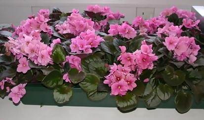
The starting point for all tissue cultures is plant tissue, called an explant. It can be initiated from any part of a plant - root, stem, petiole, leaf or flower - although the success of any one of these varies between species. It is essential that the surface of the explant is sterilised to remove all microbial contamination. Plant cell division is slow compared to the growth of bacteria and fungi, and even minor contaminants will easily over-grow the plant tissue culture. The explant is then incubated on a sterile nutrient medium to initiate the tissue culture. The composition of the growth medium is designed to both sustain the plant cells, encourage cell division, and control development of either an undifferentiated cell mass, or particular plant organs. The concentration of the growth regulators in the medium, namely auxin and cytokinin, seems to be the critical factor for determining whether a tissue culture is initiated, and how it subsequently develops. The explant should initially form a callus, from which it is possible to generate multiple embryos and then shoots, forming the basis for plant regeneration and thus the technology of micropropagation. The first stage of tissue culture initiation is vital for information on what combination of media components will give a friable, fast-growing callus, or a green chlorophyllous callus, or embryo, root or shoot formation. There is at present no way to predict the exact growth medium, and growth protocol, to generate a particular type of callus. These characteristics have to be determined through a carefully designed and observed experiment for each new plant species, and frequently also for each new variety of the species which is taken into tissue culture. The basis of the experiment will be media and protocols that give the desired effect in other plant species, and experience. The demonstration Step 1 - selection of the leaves Wash the dust off the leaves in a beaker of distilled water, holding the leaf stalk with forceps. Step 2 - surface sterilisation and preparation of the explants The leaf, with the petiole still attached, should be immersed in 70% ethanol for 30 seconds, then transferred to a sterile petri dish. Sterile scissors and forceps are then used to cut squares from the leaf as explants, each with approximately 1 cm sides. The explants are transferred into a 10% hypochlorite bleach solution for 5 minutes, gently agitating once or twice during this time. They are then washed free of bleach by immersing in four successive beakers of sterile distilled water, leaving them for 2-3 minutes in each. Three explants are placed on each petri dish of growth medium (see table and below), with the upper epidermis pressed gently against the surface of the agar to make good contact. The petri dishes are sealed with plastic film to prevent moisture loss, and incubated at 25oC in 16h light/8h dark. Step 3 - assessment of tissue culture development The media used in the demonstration are designed to show the effects of auxin, cytokinin, sucrose and mineral salts on development. The media were based on the well-known Murashige and Skoog inorganic medium, with additions as shown in this table. Typical results These pictures show typical results, after about 8 weeks on each medium. To summarise, multiple adventitious buds form on the control medium, leading to many small shoots on the upper surface where the leaf is not in contact with the medium. Absence of sucrose inhibits this production. Shoot production is also limited on the low sucrose concentration, but comparable with the control at high sucrose. At zero and low levels of cytokinin, callus forms where the leaf surface is in contact with the medium, while at high levels, shoot formation is stimulated. At zero and low levels of auxins there is a stimulus to shoot formation, but at high concentrations, large numbers of roots are formed. At low and zero levels of MS salts, there is no growth at all. These very obvious variations demonstrate the importance of a carbon and inorganic salt source for plant growth, as well as the effect of the auxin:cytokinin ration on the control of plant development.
For publications describing this work, follow this link To return to the top of this page, follow this link. |