Image Fusion for Cancer Diagnosis
A standard approach for examining excised human tissues is to stain sections with haematoxylin and eosin (H&E) and examine them with optical light microscopy. This approach results in the preservation of morphology and spatial relationships of cells and tissues and highlights the nucleic acid and protein content of the tissues at blue and red visible wavelengths, respectively. Expanding the wavelength range of tissue images to the infrared (IR) adds considerable information on the chemistry of the tissue but the longer IR wavelengths significantly reduce the spatial resolution of the image from that obtained at visible wavelengths.
Recently Safaa Al Jedani, a research student in the SciaScan group, has developed a method of fusing optical and IR images of tissue in a way that combines the spatial resolution of the optical image with the spectral information present in the IR image [1]. This insight into the chemistry of the tissue is particularly useful for histopathologist in the diagnosis of cancer. However, the technique has wider applications.
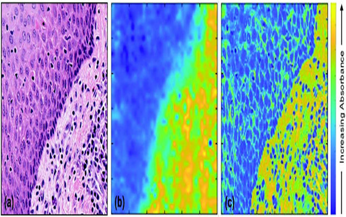
Figure (a) H&E stained image of a tissue biopsy, (b) corresponding Fourier transform IR image at 1242 cm−1 and (c) the image formed by fusing (a) and (b). While (a) shows the image at one wavelength, 1242 cm−1, there could be about 1000 fused images like (c) each obtained at a different IR wavelength and thus capturing the chemical information in the tissue.
Reference
1 "Image fusion of IR and optical microscopy for mapping of biomolecules in tissue" Safaa Al Jedani, Conor A. Whitley, Barnaby G. Ellis, Asterios Triantafyllou, Caroline I. Smith, Philip J. Gunning, Peter Gardner, Janet M. Risk, Peter Weightman and Steve D. Barrett. Analyst 146 5848 (2021).