New treatment planning approach accounting for prompt gamma range verification
In a paper recently published in the journal Physics in Medicine & Biology, OMA Fellow Liheng Tian and colleagues from Ludwig-Maximilians-Universität München have presented a new treatment planning approach accounting for prompt gamma range verification and interfractional anatomical changes.
Prompt gamma (PG) imaging is widely investigated for spot-by-spot in vivo range verification for proton therapy, however its accuracy is affected by the number of protons delivered per pencil beam and the conformity between prompt gamma and dose distribution (PG-dose correlation).
Liheng and colleagues have used a novel approach to re-optimize conventional treatment plans by boosting a few pencil beams with good PG-dose correlation above the statistics limit for reliable PG detectability. In this work, they further explore this approach with respect to the robustness of the PG-dose correlation of each pencil beam in the case of interfractional anatomical changes.
The team created a Monte Carlo treatment plan using the analytical Matlab-based treatment planning system CERR. Then they quantified the PG-dose correlation using the originally proposed approach as well as a new indicator, which accounts for the sensitivity of individual spots to heterogeneities in the 3D dose distribution. A few pencil beams were selected for each treatment field, based on their PG-dose correlation and dose surface, and then boosted in the new re-optimized treatment plan.
All treatment plans were then fully re-calculated with Monte Carlo on CT scans of the corresponding patient at three different time points (the researchers used CTs of one prostate and one head and neck cancer patient).
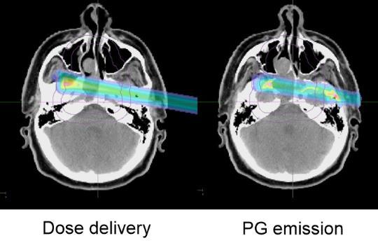
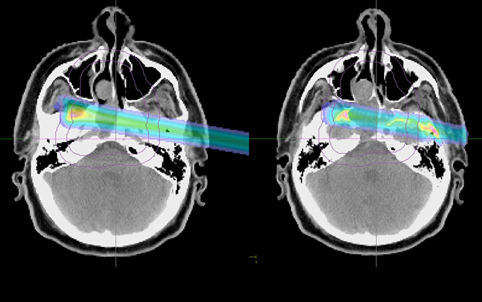
Left: Dose distribution of a given pencil beam. Right: Prompt gamma emission distribution of this pencil beam. Images courtesy of LMU.
The results show that the initial CERR Treatment Plans and the re-optimized Treatment Plans are comparable in terms of dose and dose averaged Linear Energy Transfer distribution for all CTs and patients. The recommended Pencil Beams show advantages for PG based proton range verification in terms of dose shift fidelity. Besides, compared to spot aggregation, the approach shows advantages in terms of counting statistics, lateral resolution and proton range mixing.
More information:
Liheng Tian, Guillaume Landry, George Dedes, Marco Pinto, Florian Kamp, Claus Belka and Katia Parodi, “A new treatment planning approach accounting for prompt gamma range verification and interfractional anatomical changes”, Physics in Medicine & Biology 65, 095005 (2020).