Quantification of organ motion in carbon ion therapy of abdominal lesions
Carbon ion radiotherapy (CIRT) has demonstrated to be a promising treatment for abdominal tumours, thanks to the advantages in terms of geometrical selectivity and radiobiological effectiveness with respect to X-ray and proton therapy. However, the potentially increased effectiveness and accuracy of CIRT may be undermined by the presence of organ motion. The motion of both tumour and nearby organs introduces geometric uncertainties into the process, leading to potential underdosage of the target region, and/or overdosage of organs at risk. Moreover, organ motion generates density changes within the beam path, leading to alteration in the planned dose distribution.
In a study recently published in Physica Medica, OMA Fellow Charalampos Kalantzopoulos and colleagues from CNAO and Politecnico di Milano have employed Magnetic Resonance Imaging (MRI) to quantify the tumour motion, and to evaluate the clinical approach based on the standard practice of 4D Computed Tomography (4DCT) for organ motion management.
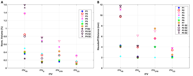
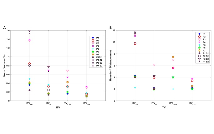
Comparison between the generated ITVs and the GTV. A) Normalized Volume and B) Hausdorff distance. *
Organ motion can be accounted for by defining margins based on the 4DCT data, with the definition of the so-called Internal Target Volume (ITV). However, 4D imaging reflects the anatomy at different time points during an “average” breathing cycle. In order to account for respiratory motion during treatment delivery, clinicians use strategies such as gating, which involves the irradiation within a particular phase of the patient’s breathing cycle.
Recently MRI has emerged as an ideal technique for time-resolved and respiratory-correlated imaging in the framework of high precision radiation therapy. MRI provides exquisite soft tissue contrast, radiation-free imaging and high temporal resolution with fast sequences. The dose-free nature of MRI and the availability of 2D fast sequences known as cineMRI, enable multiple and extended acquisitions, accounting for cycle-to-cycle breathing variations.
In this study, Charalampos and his colleagues exploited repeated cineMRI acquisition sessions to generate an ITV margin for treatment planning in CIRT of the abdominal site. Seven patients with abdominal lesions were treated with carbon-ion therapy at CNAO. The MR scan was performed on the same day of the 4DCT acquisition. For four patients, an additional MR was acquired approximately after 1 week. The cineMRI combined with deformable image registration algorithm was used to quantify tumour motion. Afterwards, two ITVs were defined considering (1) all breathing phases (ITVFB) and (2) only breathing phases within the gating window (ITVG), and then compared with the clinical (4DCT-derived) ITVs (ITVCG and ITVCFB).
The results suggest the effectiveness of the gating procedure implemented at CNAO to compensate for organ motion, and propose a method to complement the clinical workflow with cineMRI data, as a way to quantify inter and intra-fraction respiratory motion variability without delivering additional dose to the patient.
The article is part of a special issue of the European Journal of Medical Physics – Physica Medica edited on occasion of the International Conference on Medical Accelerators, organised by the OMA network in Seville (Spain) in September 2019.
The full article can be found here:
Charalampos Kalantzopoulos, Giorgia Meschini, Chiara Paganelli, Giulia Fontana, Alessandro Vai, Lorenzo Preda, Viviana Vitolo, Francesca Valvo, Guido Baroni, “Organ motion quantification and margins evaluation in carbon ion therapy of abdominal lesions”, Physica Medica 75, 33–39 (2020).
https://doi.org/10.1016/j.ejmp.2020.05.014
*image reprinted with permission from Elsevier