Studies in recognition performance reported by Vialux
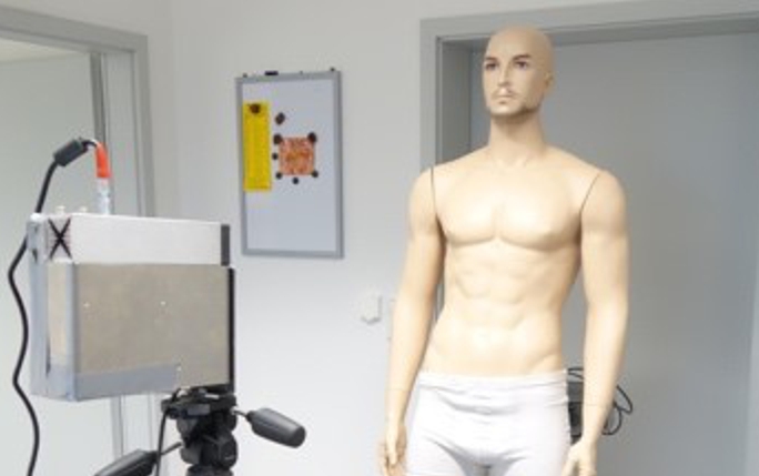
Samuele Cotta, an OMA fellow based at ViALUX, is studying the radiation hardness of ViALUX 3D scanners to be used in radiotherapy. Last September, a test with thermal and fast neutrons was performed at the FRM II research reactor in Munich. Single electronic components and a whole 3D scanner were exposed to a high neutron flux in order to reproduce in a shorter time the effects that could be observed during months of operation in a treatment room. The devices under test worked without any big issues during the irradiation and only few interruptions of the device functionality were observed and suddenly recovered through a simple reinitialization. After the tests, the irradiated 3D scanner was object of further analysis to evaluate the possible damages caused by the radiation in the CMOS image sensor, which is a key component of the ViALUX 3D scanners. The most common effect caused by the radiation is the sensor performance degradation due to the increase of the number of bright pixels, which cause a sort of fix-pattern noise in the CMOS. Several 3D images of a plastic body model were acquired to evaluate if and how these bright pixels can affect the quality of the 3D scanning process (see image above).
The acquired 3D images showed no issues at all when the typical exposure time values (5-25 ms) are used. Even with a higher exposure time (50 ms) no effect were observed (see Figure 2a) and, only increasing it up to 250 ms, few missing 3D points were observed (see image below).
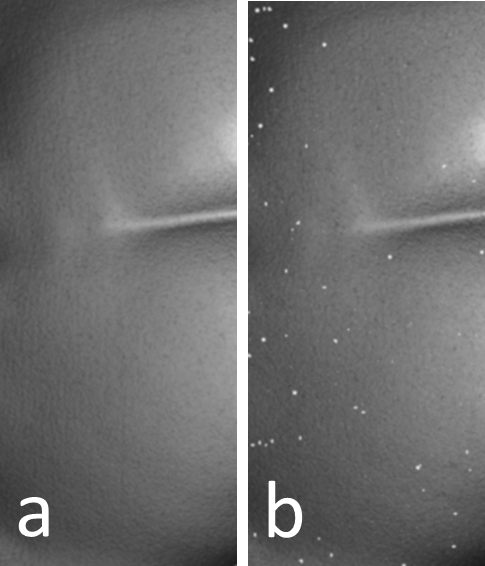
Detail of a 3D image of the body model acquired with an exposure time of 50 ms (a) and 250 ms (b).
Samuele is now considering possible solutions to further improve the radiation hardness of the device, even if the tests have already given a positive feedback about the reliability of the ViALUX devices.