Design and optimisation of ultra-compact, high resolution X-ray imaging systems
To study the optimisation of X-ray imaging systems and to improve the diagnostic arsenal of clinical practice, a collaboration between the University of Liverpool and Adaptix Ltd. has been funded by the Science and Technology Facilities Council under the Accelerators for Safety, Healthcare and Environment program.
The idea is to design an ultra compact and high resolution digital tomosynthesis system which shall be the intermediate step between the conventional and low dose planar X-ray and the state of the art but high dose and expensive computerised tomography, giving doctors the flexibility to choose a tool that combines both 3D and low dose.
Digital tomosynthesis is an old technology that got overshadowed by computerised tomography since the latter creates better images. However, decades of use and research revealed the detrimental effect of the high doses from computerised tomography and an alternative 3D technology with much lower dose is desired. So digital tomosynthesis is now optimised to bridge this gap. Digital tomosynthesis works by moving the X-ray source around the patient and acquiring images like computerised tomography but instead of a full 360 degree rotation, the angle is much smaller. This reduces the dose but the smaller angle means less information is used for the reconstruction leading to lower image quality and moving the source is also responsible for motion induced artefacts. To tackle this, the designs include a system with multiple sources at different positions and angles that replace the one moving source. This minimises motion induced artefacts but creates new challenges that need to be investigated.
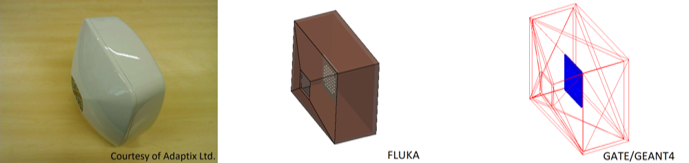
The images show the geometry of the system in reality and partly constructed in the two simulation programs FLUKA and GATE/GEANT4.
This research uses Monte Carlo simulations to study how the system designs effect its performance and finally optimises the designs according to the resulting performance.
The study includes among others:
• electron acceleration
• X-ray generation, filtration and collimation
• patient dose
• detector response
Therefore, the simulation framework is both complete and transparent and can be tested on different Monte Carlo simulation codes. Benchmarking with experimental results gives confidence on the simulations and transparency allows benchmarking to be done at the different levels of the study.
Currently involved QUASARs:
Thomas, Carsten