Physics research at the University of Liverpool

Nanoscience

The overall scope of the research programme is the exploration of the fundamental physics underlying and underpinning developments in a range of applied areas including nanotechnology, sensor development, clean energy sources and environmentally friendly materials.

Dr Steve Barrett Department of Physics
Dr Yvonne Grunder Department of Physics, Stephenson Institute for Renewable Energy
Dr Frank Jaeckel Department of Physics, Stephenson Institute for Renewable Energy
Professor Chris Lucas

Department of Physics, Stephenson Institute for Renewable Energy

Professor Ronan McGrath

Department of Physics, Surface Science Research Centre

Dr Hem Raj Sharma

Department of Physics, Surface Science Research Centre


Electrochemical cell for in-situ X-Ray Scattering studies

In-situ X-Ray Scattering Studies of Electrochemical Systems


In the last decade, synchrotron surface x-ray diffraction has been a critical tool for determining the potential dependence and stability of specific surface structures in electrolyte under reaction conditions. In-situ diffraction is a unique tool that allows structural characterisation of the interfacial atomic structure including subsurface and ordering on the electrolyte side of the interface.


Read more...

We aim to understand the structure-function relationship of the electrochemical interface, thus investigating structures of metal electrodes in-situ gives unique information how changes in structure and morphology affect electrochemical properties. In addition through the structural characterisation, bond lengths can be extracted which give indirect information about the chemical nature of surface and ad-atoms.

Studies have been made of systems relevant to electrocatalysis, batteries and electroplating processes. The experiments involve physical phenomena such as surface reconstruction, atomic layer deposition, surface phase transitions, surface segregation and chemical reactions.

We recently extended the in-situ characterisation to resonant surface x-ray diffraction that gives direct information about the chemical properties (e.g. charge distribution). Experiments are conducted by scanning the x-ray energy of the incident beam through the edge of an element present at the electrode while recording the diffracted intensity at a key point in reciprocal space.

The next stage in this project is to further investigate temperature effects in electrochemistry (link to the other section) and to extend our studies to non-aqueous electrolytes.

Experiments are carried out at the EPSRC mid-range facility beamline, XMaS, at the ESRF but also at other beamlines at the ESRF, the APS, the SLS and the Diamond Light Source.

For further information watch the video of Professor Chris Lucas talking about Structure at the Electrochemical Interface.

Personnel:  Dr Yvonne Grunder, Professor Christopher Lucas

Electrochemical characterisation of solid-liquid interfaces


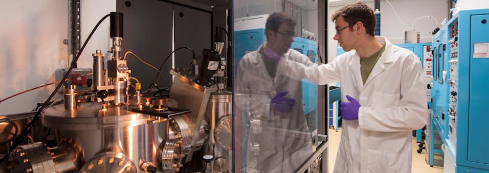
Electrochemical investigation is carried out in our new electrochemical laboratories in the Stephenson Institute for Renewable Energy. We currently have a couple of measuring stations allowing the study of electrochemical surface processes by electrochemical methods (e.g. cyclic voltammetry, chronoamperometry).


Read more...

We are currently developing a UHV-electrochemical transfer cell (in collaboration with Dr Vin Dhanak), which will allow the transfer of electrodes of more reactive materials from a UHV preparation chamber to an electrochemical cell (either mounted on the UHV chamber or situated in a glove box) without exposure to ambient. This will also enable the study of ionic liquid electrolytes in a vacuum environment.

In addition a glove box will soon be acquired for our studies of electrochemistry in non-aqueous electrolytes allowing the characterisation of the systems in controlled atmospheres.

Current projects include:

  • Underpotential deposition
  • Adlayer formation
  • Electrocatalysis
  • Temperature effects
  • Non-aqueous electrolyte solutions

Personnel:  Dr Yvonne Grunder, Professor Christopher Lucas

Epitaxy on Quasicrystal Surfaces and Intermetallics in Catalysis


Quasicrystals are new type of solids that differ from other two known forms, crystal and amorphous, by possessing long range order without periodicity. They are of specific stoichiometry and often exhibit crystallographically forbidden rotational symmetries such as tenfold and fivefold.


Read more...

We have shown using scanning tunnelling microcopy (STM) that despite their complex aperiodic structures, quasicrystals and related periodic approximants yield atomically flat surfaces characteristic of bulk truncations. We have then explored the modification of their structure using adsorbates and delineated the conditions under which ordered adsorption can take place.

We also study surfaces of intermetallics that are potential to be used as catalysts in steam reforming of methanol. The steam reforming is one of the most promising processes to provide hydrogen for mobile fuel cell. The intermetallics of current investigation include ZnPd alloys and quasicrystals. Most quasicrystals contain catalytically active elements and they are brittle to facilitate high surface areas through crushing.

We have recently reported the first examples of template quasicrystalline ordering of a single element (Nature Communications 2013) and molecules (Nano Letters 2014). We are involved with the European research network for intermetallic compounds as catalysts for steam reforming of methanol (COST Action CM0904) and the European Integrated Centre for the Development of New Metallic Alloys and Compounds.

Personnel:  Professor Ronan McGrath, Dr Hem Raj Sharma

Transmission electron micrograph of colloidal quantum dots.

Hybrid Nanomaterials


Nanomaterials exhibit properties significantly different from their bulk counterparts due to quantum confinement effects. This allows tuning their electronic and optical properties beyond what is possible in the bulk.


Read more...

Hybrid nanomaterials on the other hand combine different material classes such as metals, semiconductors or organic materials on the nanoscale. In addition to quantum confinement effects, this can give rise to enhanced or novel properties that none of the individual components exhibit. Such properties include but are not limited to fluorescence, Raman and Rayleigh scattering, chiroptical properties, and catalytic activities. We are interested in the preparation and advanced optical characterisation of hybrid nanomaterials for applications in nanoscale optics, plasmonics, photonics and renewable energy.

Personnel:  Dr Frank Jaeckel

Scanning tunnelling microscope image of surface of scandium

Scanning Microscopy and Electron Diffraction


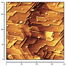
Scanning microscopy techniques are used extensively in nanoscience. Scanning Probe Microscopy (SPM) is a generic term encompassing Scanning Tunnelling Microscopy (STM), Atomic Force Microscopy (AFM), Scanning Near-Field Optical Microscopy (SNOM) and related techniques. In addition, Scanning Auger Microscopy (SAM), Scanning Electron Microscopy (SEM) and Transmission Electron Microscopy (TEM) are often used in nanoscale characterisation of materials. Images generated from all of these microscopy techniques need processing and analysis to extract quantitative information.


Read more...

The images produced by these techniques share a number of related features: (i) the image files have a file format that is specific to the manufacturer of the microscope system, and (ii) the image files contain associated acquisition parameters, such as spatial calibration data for the x and y (and sometimes z) axes. If a user wishes to carry out off-line processing or analysis of their images using software other than that provided by the manufacturer of their microscope system, then these features can be a potential problem.

Image SXM is image analysis software that has been extended for use with scanning microscope images. Examples of how it has been used include molecular shape recognition and identifying molecular registration.

A complementary technique to SPM in determining surface structure involves analysis of the diffraction patterns produced in low-energy electron diffraction (LEED). Quantitative analysis of the variation of diffraction spot intensities (I) as a function of electron beam energy (or acceleration voltage, V) is referred to as LEED I-V. Comparison of experimental I-V spectra with theoretical spectra derived from models of surface structures can be used to determine atomic positions to very high precision. Image SXM can extract LEED spot intensities automatically from a set of LEED images or a video of the LEED pattern varying as the accelerating electron voltage is ramped. For an example of how this has been used in surface structure determination, see J. Chem. Phys. (2005).

Personnel:  Dr Steve Barrett

Back to: Department of Physics