Biophysics uses the quantitative methodology of physics to study biological systems. Biophysics research spans spatial scales ranging from the molecular level (nm), through cellular structure (nm to µm), to the structures of tissues (µm to mm). By its very nature it is an interdisciplinary science, and the Biophysics Research Cluster comprises members of the Department of Physics and staff from other departments with overlapping interests:
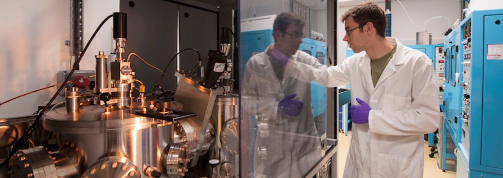
Microscopy techniques, such as atomic force microscopy (AFM) and scanning near-field optical microscopy (SNOM), are applied across all scales from nm to mm and the development of image processing and image analysis techniques is an integral part of the research.
Molecular Organisation and Dynamics
Microscopy and spectroscopy techniques are used to study how molecules organise themselves and how they move.
Reflection anisotropy spectroscopy (RAS) is used to monitor the organisation and dynamics of molecules adsorbed on surfaces by (i) revealing the three dimensional orientation of molecules adsorbed at metal liquid interfaces; (ii) distinguishing between single and double stranded DNA adsorbed on surfaces, showing the potential as a fast cheap and accurate method of genetic screening; and (iii) monitoring conformational change in proteins arising from electron transfer in real time.
The macroscopic structures of tissues, both healthy and diseased, are studied to improve our understanding of their function.
SpectroChemical Analysis for Cancer (SCAnCan) is an EPSRC-funded programme to advance the understanding of oesophageal, cervical and prostate cancers through the application of infrared, Raman and THz techniques. A key role is played by equipment the group have constructed on the ALICE accelerator, the UK’s only fourth-generation light source and free electron laser, located at Daresbury Laboratory. Various imaging techniques are being used in an attempt to identify the characteristic signatures of pre-cancerous tissues in infrared absorption at selected wavelengths. Scanning near-field optical microscopy (SNOM) provides images that beat the diffraction limit and give spatial resolutions of significantly less than the wavelength of the infrared light. Preliminary results have been presented in Appl. Phys. Lett. (2013) and the programme was described on BBC Today in February 2012.
Bioartificial tendons are used as models to understand the structure and biomechanics of, and the initiation of microdamage in, musculoskeletal tissues.
The structure of the sclera of porcine eyes is being studied using atomic force microscopy (AFM), transmission electron microscopy (TEM) and polarised light microscopy (PLM). Image analysis techniques are being developed to quantify the structures imaged.
Image analysis techniques are used to allow quantitative data to be extracted from all types of microscopy used in biophysics and related fields.
Image SXM is the general purpose image analysis software that has been extended to produce customised solutions for a number of medical applications, collectively labelled MIASMA. These include:
MIASMA project
Relevance to
Collaborator
Carbon particulates
Lung cancer
Dr Stephen Gordon
Parasite analysis
Malaria
Prof. Alister Craig
Capillary blood flow
Meningitis
Dr Richard Sarginson
Retinal analysis
Diabetes
Dr Yalin Zheng
Parasite morphology
Leishmaniasis
Dr Rod Dillon
Assessing antibiotics
Tuberculosis
Dr Derek Sloan
Fibrillin microfibrils
-
Dr Riaz Akhtar
Microcompartments
-
Dr Luning Liu
In addition, image analysis is playing a key data analysis role in the major project SpectroChemical Analysis for Cancer (SCAnCan) described in the previous section.