Histology Facility
Providing a co-ordinated and unified approach to cover the entire histological process
Providing a co-ordinated and unified approach to cover the entire histological process
The newly established Histology Shared Research Facility encompasses two well equipped laboratories that offer researchers the full histological process from preparing your samples through to scanning images.
The equipment we offer includes:
Our specialist technical staff can prepare your samples from fresh or fixed tissue through to scanned images that we can then transfer to you for analysis of your research.
We can train staff and students to prepare and use our equipment to embed, section and hand stain your tissues as required. We currently offer three different types of wax for embedding and sectioning as required.
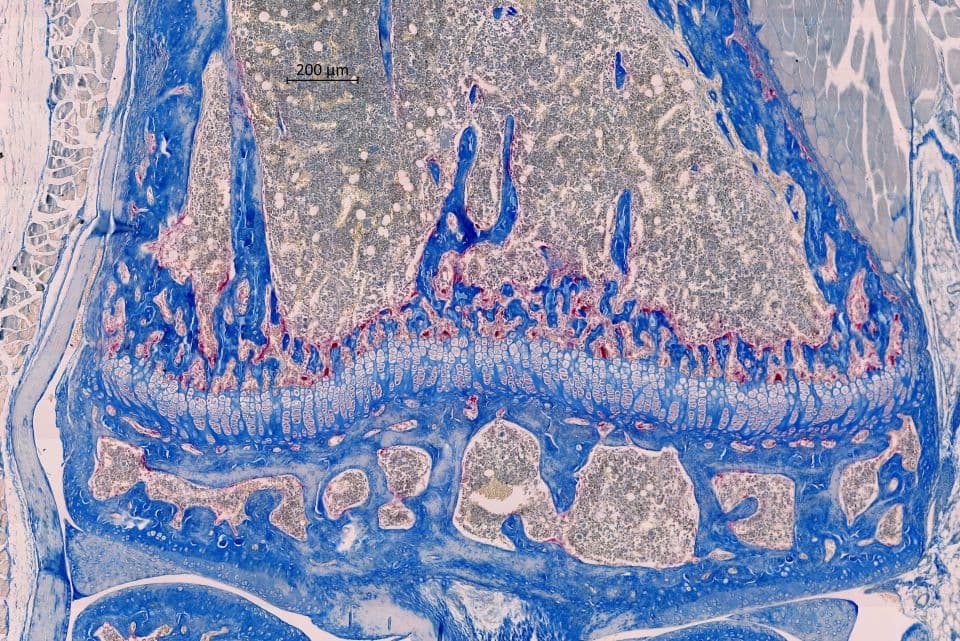
We are fully equipped for methyl methacrylate and glycol methacrylate embedding and sectioning (including larger samples), freeze microtomy and high throughput slide imaging using our Zeiss Axioscan Z1.
Back to: Research
Booking duration and rates for facilities and equipment can be flexible depending on your research needs.
Please email us on Histology@liverpool.ac.uk to discuss your research.
Automated machines that dehydrate tissue, under vacuum, in preparation for embedding in wax or methacrylate.
Contains a reservoir of molten wax ready to embed tissues. We have three different waxes for specific requirements.
For tissue sectioning, both automated and fully manual for sectioning of paraffin wax and methacrylate embedded samples.
For sectioning of frozen tissue.
For cutting accurate semi-thin sections of delicate fresh tissue.
Microtome for cutting accurate semi thin sections of frozen tissue.
For cutting accurate thin sections of larger methacrylate embedded tissue samples.
For imaging brightfield and fluorescent stained slides.

Histology technician