Work Experience Students’ Photomicrographs

Fay, Dylan and Dan are Year 10 students who worked in the CTL as part of their work experience placement at the University of Liverpool. During their time in the CTL they used different microscopes with camera attachments and took photographs of thin sections of rocks, micro-organisms and fossils. Each student chose one of the images they produced and wrote a brief description of what their image showed.
Faye Mills, Calderstones School

In this image I captured is an example of a limestone. Limestone is a sedimentary rock composed of calcium carbonate (CaCO3). The coloured dye at the top is an indicator of iron (Fe) in the calcium carbonate. (Note: The blue colour indicates the presence of iron)
Dylan Owen, Wirral Grammar School for Boys
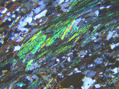
This example of mica crystals in schist is a good example of foliation (the parallel alignment of minerals in a metamorphic rock). These minerals are green at this angle though in different angles they refract light into different colours.
Dan Tomlinson, Stanley High School
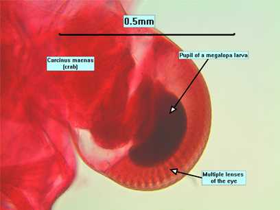
It shows an in-detail zoom image of the eye of a tiny megalopa larva. It has multiple lenses in its eye. The image also has a scale to show how small the larva is.